All images on this website have been taken in Leicestershire and Rutland by NatureSpot members. We welcome new contributions - just register and use the Submit Records form to post your photos. Click on any image below to visit the species page. The RED / AMBER / GREEN dots indicate how easy it is to identify the species - see our Identification Difficulty page for more information. A coloured rating followed by an exclamation mark denotes that different ID difficulties apply to either males and females or to the larvae - see the species page for more detail.
Fungi
Fungi are not plants, as was thought to be so in the past, but in a separate Kingdom of their own. In most cases, the main body of the fungus is hidden from view. It is a network of threads (hyphae) collectively called a 'mycelium', which permeate the substrate on which the fungus grows. The hyphae absorb nutrients and water from the substrate. The reproductive spore-producing structures, such as the familiar mushroom or bracket, are the visible parts of the fungus.
The Fungus Kingdom is extremely diverse, and includes many microfungi that are rarely recorded. Most of the larger species are in one of two major divisions or Phylum, based on the way the fungus reproduces. Basidiomycota (sometimes called 'spore-droppers') include mushrooms and toadstools, brackets, corals, puffballs, jellies, rusts and smuts. The Ascomycota or 'spore-shooters' includes cups, morels, yeasts and many other smaller fungi.
Taxonomy is very complicated and is liable to change as the true relationships between species are worked out. In the sections below, we have grouped fungi into categories based on their structure and appearance rather than their place in the scientific taxonomic hierarchy. There is more information under each section.
Recording fungi
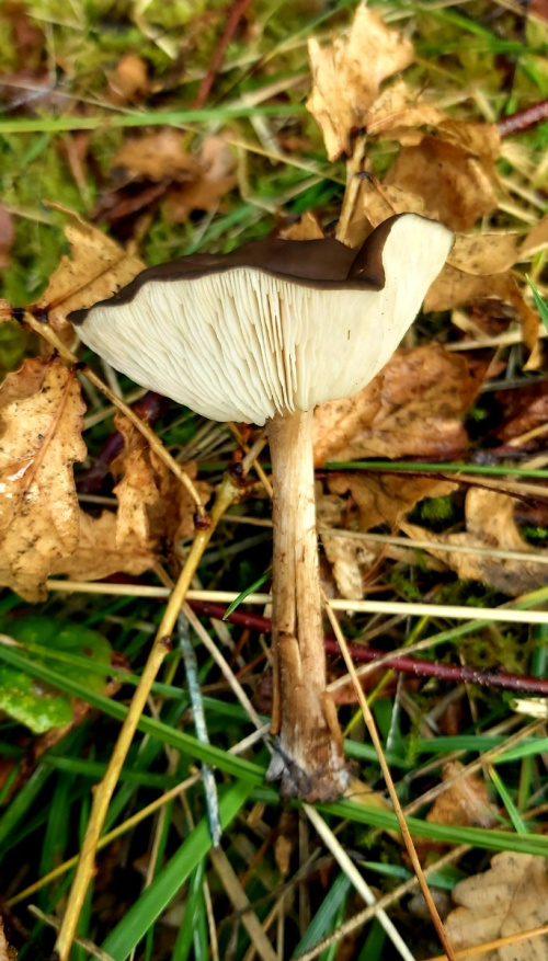
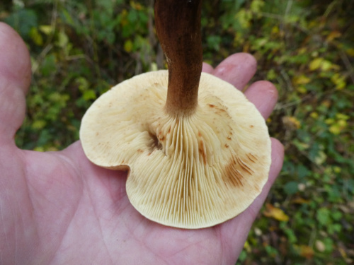
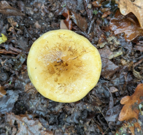
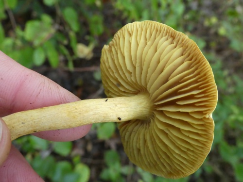
Always note the substrate or host-plant on which the fungus is growing, and the habitat. To identify many large species, it is helpful to get a good photograph of the cap (from the top), the gills or spore-producing structures (from underneath) and the stipe or stem (from the side) - three images from three angles, rather than images from much the same angle.
Always note the texture of cap and stipe; the smell; and the presence of any staining or 'milk' produced when the cap, gills, stipe or body of the fungus is bruised.
The colour of the spores can be very helpful. It is best seen by taking a spore-print from a collected specimen - advice on doing this is in many of the resources below. For some fungi, microscopic examination of the spores may be needed for identification.
Resources
The Leicestershire Fungi Study Group is a great place to start if you want to learn about fungi. They welcome new members. Their annual programme of study forays is led by enthusiastic and knowledgeable leaders. There are also indoor microscopy sessions held in Leicester. This is a really excellent way to broaden your knowledge, and make friends who share your interest in fungi.
There are many field guides, but none of them contain all the fungi you will find, or have all the photos or information you need, so it is a good idea to check several different books and websites.
- Kibby, G. 2020-2022. Mushrooms and Toadstools of Britain & Europe, Volumes 1 - 4. (privately published)
- Laessoe,T. & Petersen,J. (2019) Fungi of Temperate Europe. Princeton University Press, Volumes 1 and 2
- O'Reilly, P. (2016). Fascinated by Fungi. First Nature
- Sterry, P. & Hughes, B. (2009). Complete guide to British Mushrooms and Toadstools. Collins
- Wood, E & Dunkelman, J. (2017). Grassland Fungi: a Field Guide. Monmouthshire Meadows Group
Useful websites include Pat O'Reilly's First Nature website, the Buckinghamshire Fungus Group, the Hampshire Fungus Recording Group and Fungi Outer Hebrides.
The British Mycological Society website is a good source of information, mostly aimed at the expert.
If you know of other websites or books that you would recommend, do let us know: info@naturespot.org
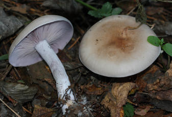
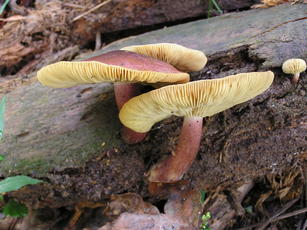
Agarics and other fungi with caps and gills
In this section are most of the commonly recorded 'mushrooms and toadstools' with spore bearing gills underneath the cap. Most of these are Agarics and related species, but we have included some families that have caps and gills but are unrelated - e.g. the Russulaceae.
For most species, the spores fall from the gills and are dispersed by the wind. Spore colour is an important way of identifying species and it may be necessary to take a spore print from a collected specimen. As well as noting the colour, smell, texture, milk/staining, habitat and substrate, also note:
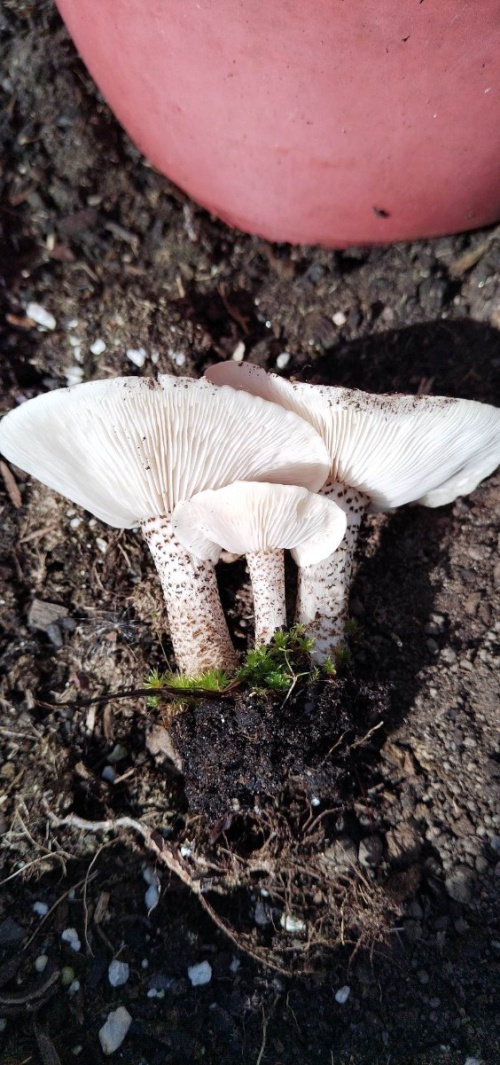
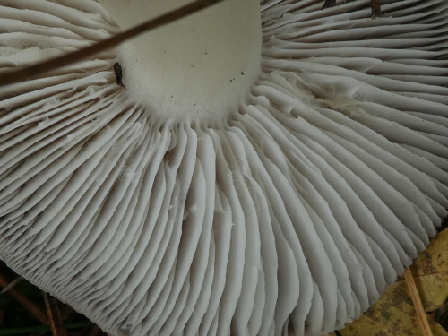
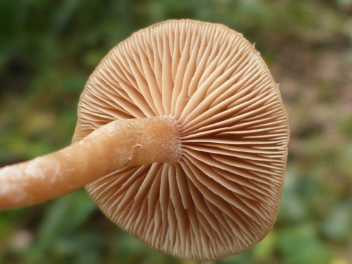
- the way the gills are attached to the stem or stipe; cutting vertically through the cap and stipe will help to see this (some examples are here: decurrent; adnate; adnexed/free)
- the colour of the gill edges (example: velvet shield)
- whether there is a ring around the stipe (some examples: wood mushroom; porcelain fungus)
- whether there is a volva or sac at the base of the stipe (example: false death-cap)
- whether there is a veil or the remains of one under the cap. (example: birch web-cap
Tricholomataceae - Knights, Cavaliers, Funnels, Blewits, etc.
A large family that includes the following important genus - at least currently (2026); it may be subject to change:
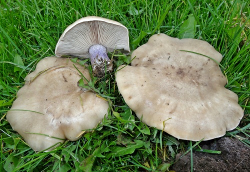
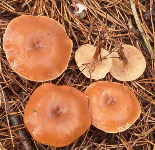
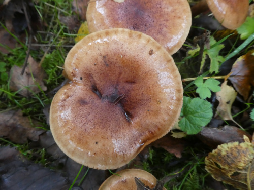
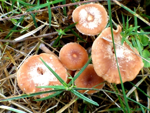
Funnels (Clitocybe and Infundibulicybe) have white or cream or pinkish-clay spore-prints. It can assist identification to take a print from a specimen; spore size may also be important. Not all are funnel shaped but when mature usually have a depressed centre and gills that are decurrent, sometimes strongly so. Smell should also be noted; e.g. some smell farinaceous (i.e. floury); others have sweet, spicy or aniseed smells. In the gallery below, and exclamation mark after the camera icon indicates that a spore-print will be helpful or that a distinctive smell should be noted.
Blewits (Lepista) are medium to large fungi, most with pinkish or brownish-pink warty spores, and often with violet or lilac tones and a pleasant flowery scent.
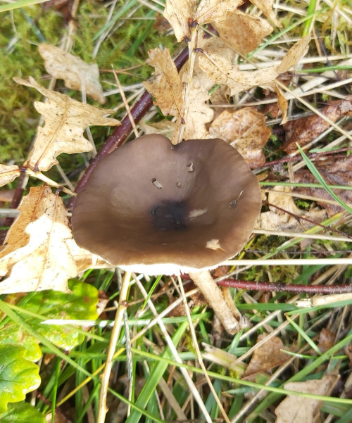
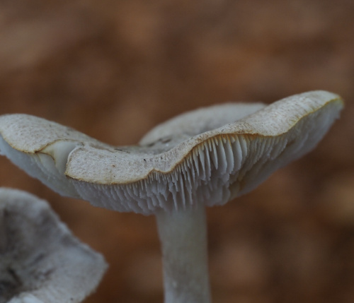
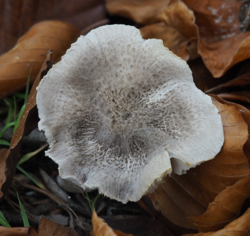
Cavaliers (Melanoleuca) are a large and difficult genus, mostly with flattened or umbonate caps and white warty spores.
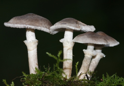
Knights (Tricholoma) are another large and difficult genus. They have white smooth spores and usually have emarginate gills; some have a fine veil, the remains of which can persist as a ring on the stipe. It is always helpful to sniff the fresh specimens; some have distinctive (or no) smells.
In the galleries below, an exclamation mark after the red or amber camera icon means that microscopic examination or information on smell, taste, texture or another factor is needed.
[NB: We have included the Funnels and Blewits (Clitocybe, Infundibulicybe, Lepista and other related species) here because they are placed in Tricholomataceae on the BMS GB checklist - however, these species are currently 'Incertae sedis' in the Agaricales on the NBN (i.e. not assigned to any Family). A separate family, Clitocybaceae, is widely used, as on CABI database.]


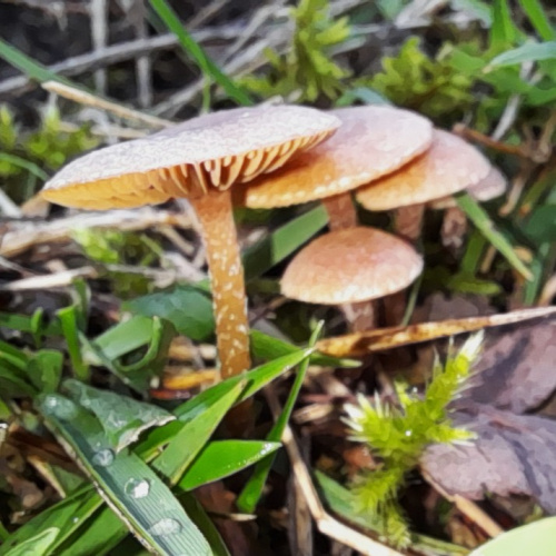
Tubariaceae - Twiglets
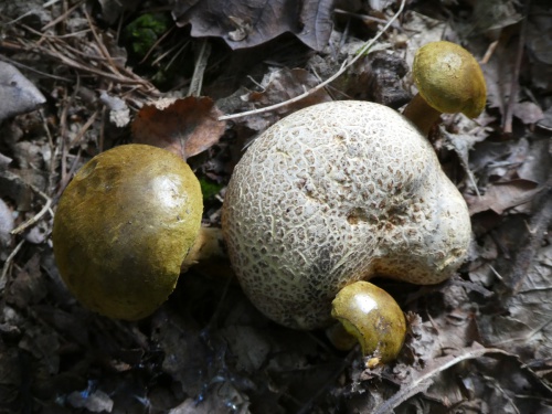
Boletes: fungi that have caps with pores
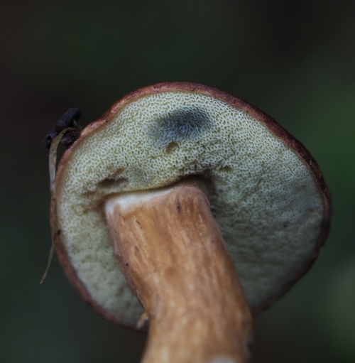
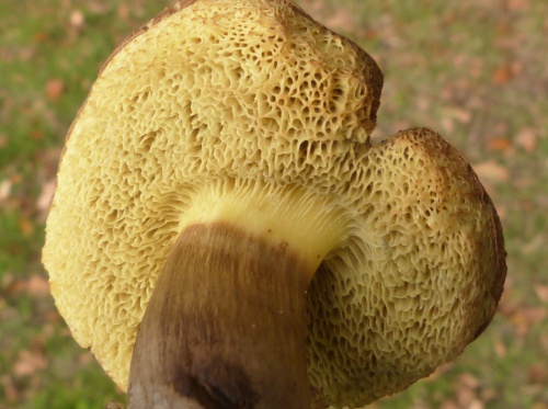
These fungi have spongy tissue made up of long narrow tubes ending in pores on the underside of the caps. Unlike some stemmed polypores (see Lentinus, etc.) the tubes can easily be detached from the flesh. The spores are usually olive-green or brown, and drop out from the pores.
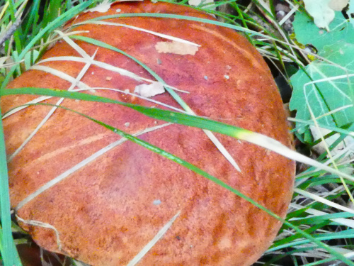
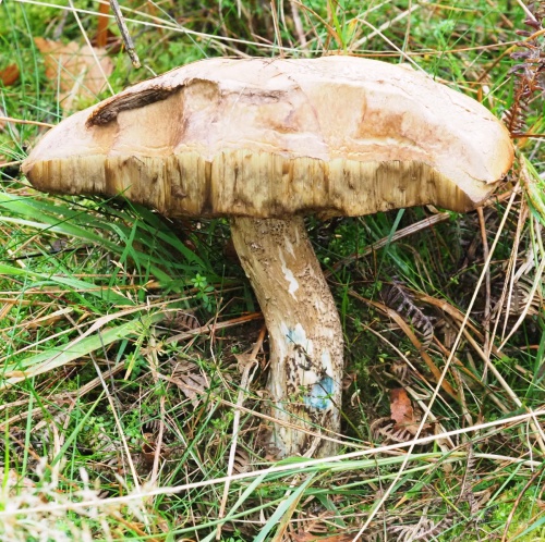
- Photograph a fresh specimen from top down, in side view and underneath to show pores and full length of stem.
- Don't try and identify one that is over-mature, or has been over-nibbled by slugs or is infested with maggots.
- Substrate and plant associations are important; always note the tree species if it is found under trees
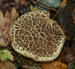
- Photograph or note the texture of cap (e.g. felted, slimy, sticky, scaly, cracking) and note or photograph any colour change below the outer cap surface (the subcuticular layer) - e.g. where is has been nibbled.
- Note the size and colour of the pores
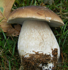
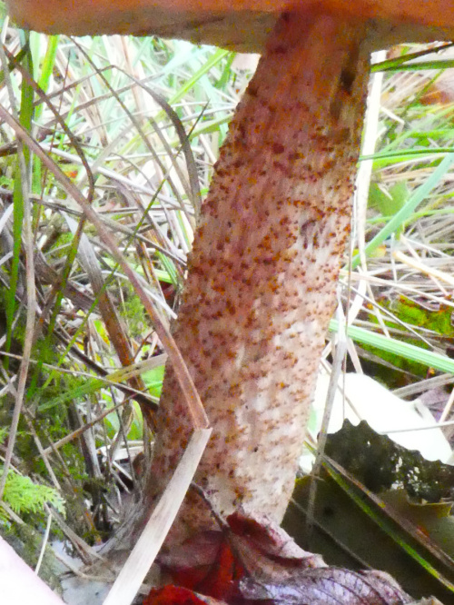
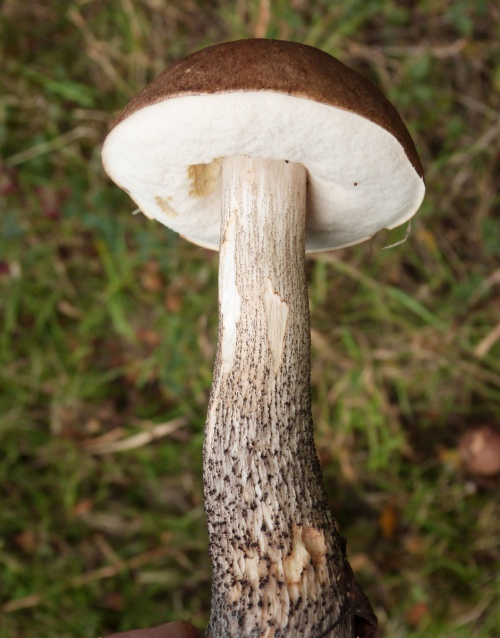
- Photograph or note the detailed surface texture of the stipe, which may be smooth or covered in a fine net or reticulation (see cep) dots or scales (see orange birch bolete).
- Photograph or note whether a stem ring is present (as in some Suillus species e.g. the Larch bolete)
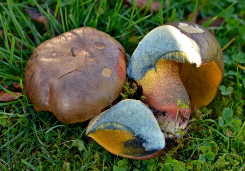
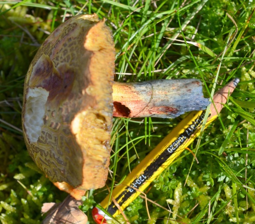
- Many species change colour when bruised (example: blue coloration on bruised pores of bay bolete). It is helpful to slice a specimen vertically through the cap and stipe to show the flesh colour and areas of staining. Note colour of flesh and any flushed areas of a different colour; note any blue-green stains and whether this developes slowly or quickly; note which part of the specimen (e.g cap flesh, top or base of stipe) the colour change is seen.
There is more information in Kibby, G. ( 2017) British Boletes, with keys to species (8th edn., privately publ.). Verification from photos can be difficult - colours and textures do not always transfer well - and we recommend that identification is checked from a specimen, preferably by following a key or with expert advice. Taxonomy can be confusing; some species appear in various sources under a range of dfferent names. In the gallery below, we have followed names on the National Biodiversity Network (NBN) in January 2026, but have added synonyms to the species pages.
Some related fungi have gills rather than pores - e.g. the Roll-rims, False Chanterelle and Spikes - so we have included these in the Gill Fungi section (see Paxillaceae, Hygrophoropsidaceae and Gomphidiaceae).
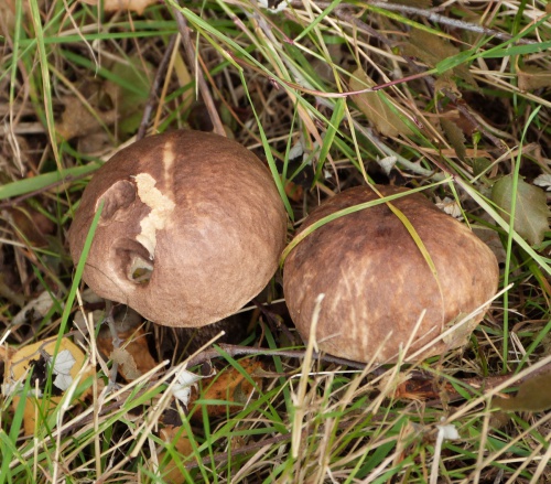
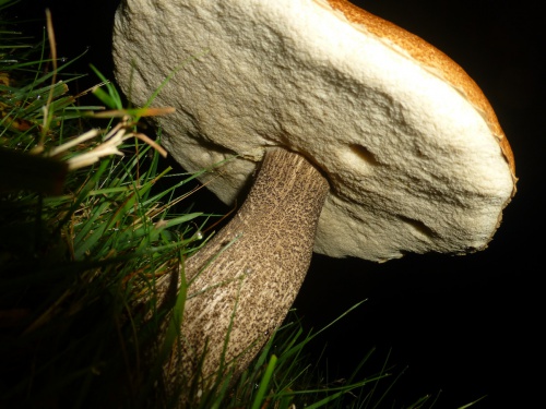
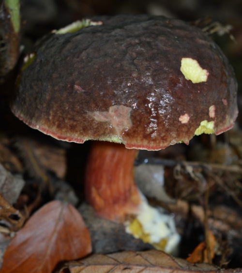
Boletaceae - Boletes
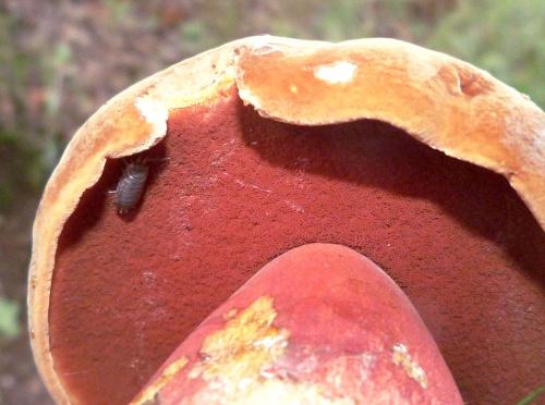
![]() grey-brown fibrillose stipe
grey-brown fibrillose stipe
yellow apex and narrow red zone below
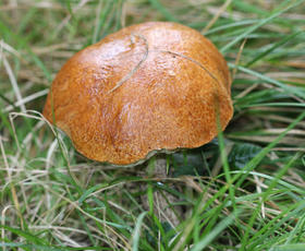

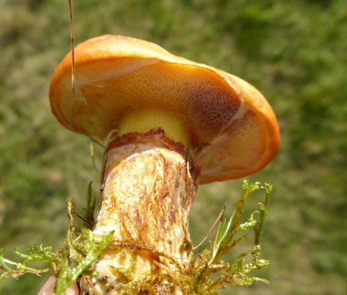
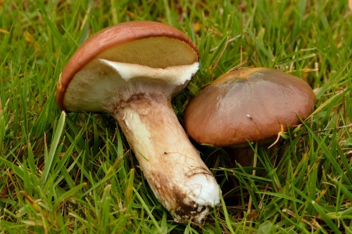
Suillaceae - Boletes
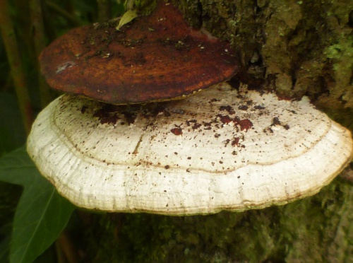
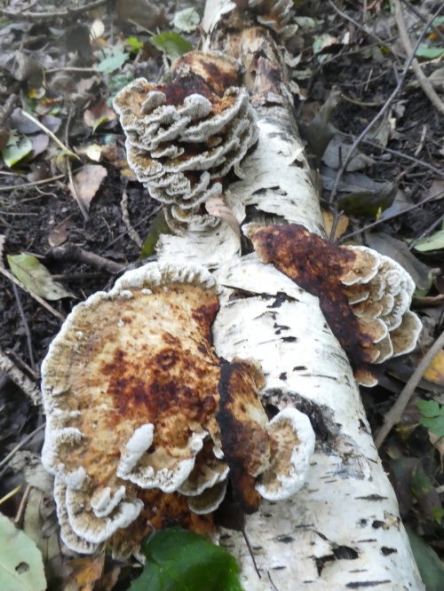
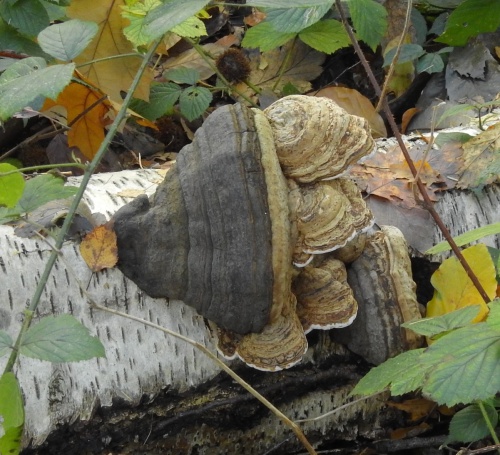
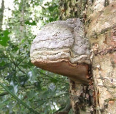
Polypores and Corticoids - brackets and crusts
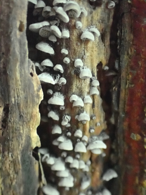
This section includes many of the fungi that grow on the trunks, branches and stumps of trees. Often the species of tree on which they are growing - including dead wood - is an aid to identification. Almost all are decomposers of wood, causing white or brown rot.
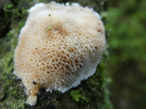
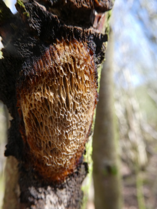
Polypores release spores through downward pointing tubes on fertile surface. Pores can be labyrinthine (maze-like - see Oak Mazegill), circular, angular (e.g. Alder Bracket), elongated (e.g. Lumpy Bracket) or stretched and gill-like; always examine and photograph the pore layer. The number of pores per millimetre can be an important feature (use a transparent rule and hand lens). Some pore surfaces stain when bruised. Polypores can be perennial or annual; perennials e.g Artist's Bracket have several tube layers and often have zonate caps and hard or tough fruitbodies; annuals can be soft or tough and leathery. Polypores can have several different growth forms:
- Capped - a shelf/bracket or hoof-like form with a distinct cap on upper surface and a fertile surface with pores underneath e.g. Blushing Bracket or Hoof Fungus
- Resupinate – a flattened fruitbody without a cap, appressed to the substrate (e.g. Rusty Porecrust);
- Reflexed – partly resupinate, partly capped, with part of the fruit body bent back to form a bracket-like cap. e.g. Smoky Bracket;
- Clustered or fasciculate - numerous caps from a narrow common base or short thick stem e.g. Chicken-of-the-Woods, Dyer's Mazegill.
- Stemmed - tongue shaped with lateral stem attached to substrate (e.g. Birch Polypore or Dryad's Saddle), or with a central or acentric stem (e.g Winter Polypore and Bay Polpore). The latter may be superficially similar to Boletes, but polypore tubes cannot be separated from the cap flesh, unlike boletes
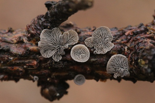
Corticoids do not have pores on the fertile surface and are usually resupinate or reflexed but may also be mould-like or fragile and loosely attached. Always photograph the fertile layer as well as the cap of reflexed forms. Some fertile surfaces stain when bruised e.g. Bleeding Oak Crust. The fertile surfaces can be wrinkled (merulioid) e.g. Wrinkled Crust or Netted Crust; smooth e.g. Hairy Curtain Crust, spiny or toothed e.g. Toothed Crust, gill-like e.g. Crimped Gill, or warty. Most are annual. Identification of many species is very difficult, and we have only illustrated a small number of UK species below. Microscopic examination by an expert will be needed to identify some.