All images on this website have been taken in Leicestershire and Rutland by NatureSpot members. We welcome new contributions - just register and use the Submit Records form to post your photos. Click on any image below to visit the species page. The RED / AMBER / GREEN dots indicate how easy it is to identify the species - see our Identification Difficulty page for more information. A coloured rating followed by an exclamation mark denotes that different ID difficulties apply to either males and females or to the larvae - see the species page for more detail.
Fungi
Fungi are not plants, as was thought to be so in the past, but in a separate Kingdom of their own. In most cases, the main body of the fungus is hidden from view. It is a network of threads (hyphae) collectively called a 'mycelium', which permeate the substrate on which the fungus grows. The hyphae absorb nutrients and water from the substrate. The reproductive spore-producing structures, such as the familiar mushroom or bracket, are the visible parts of the fungus.
The Fungus Kingdom is extremely diverse, and includes many microfungi that are rarely recorded. Most of the larger species are in one of two major divisions or Phylum, based on the way the fungus reproduces. Basidiomycota (sometimes called 'spore-droppers') include mushrooms and toadstools, brackets, corals, puffballs, jellies, rusts and smuts. The Ascomycota or 'spore-shooters' includes cups, morels, yeasts and many other smaller fungi.
Taxonomy is very complicated and is liable to change as the true relationships between species are worked out. In the sections below, we have grouped fungi into categories based on their structure and appearance rather than their place in the scientific taxonomic hierarchy. There is more information under each section.
Recording fungi
Always note the substrate or host-plant on which the fungus is growing, and the habitat. To identify many large species, it is helpful to get a good photograph of the cap (from the top), the gills or spore-producing structures (from underneath) and the stipe or stem (from the side) - three images from three angles, rather than images from much the same angle.
Always note the texture of cap and stipe; the smell; and the presence of any staining or 'milk' produced when the cap, gills, stipe or body of the fungus is bruised.
The colour of the spores can be very helpful. It is best seen by taking a spore-print from a collected specimen - advice on doing this is in many of the resources below. For some fungi, microscopic examination of the spores may be needed for identification.
Resources
The Leicestershire Fungi Study Group is a great place to start if you want to learn about fungi. They welcome new members. Their annual programme of study forays is led by enthusiastic and knowledgeable leaders. There are also indoor microscopy sessions held in Leicester. This is a really excellent way to broaden your knowledge, and make friends who share your interest in fungi.
There are many field guides, but none of them contain all the fungi you will find, or have all the photos or information you need, so it is a good idea to check several different books and websites.
- Kibby, G. 2020-2022. Mushrooms and Toadstools of Britain & Europe, Volumes 1 - 4. (privately published)
- Laessoe,T. & Petersen,J. (2019) Fungi of Temperate Europe. Princeton University Press, Volumes 1 and 2
- O'Reilly, P. (2016). Fascinated by Fungi. First Nature
- Sterry, P. & Hughes, B. (2009). Complete guide to British Mushrooms and Toadstools. Collins
- Wood, E & Dunkelman, J. (2017). Grassland Fungi: a Field Guide. Monmouthshire Meadows Group
Useful websites include Pat O'Reilly's First Nature website, the Buckinghamshire Fungus Group, the Hampshire Fungus Recording Group and Fungi Outer Hebrides.
The British Mycological Society website is a good source of information, mostly aimed at the expert.
If you know of other websites or books that you would recommend, do let us know: [email protected]
Clubs, Corals and Spindles
In this section we have grouped species with club-shaped, awl-shaped or branched fruit-bodies. All are basidiomycetes, but this is an artificial grouping - despite appearances, they are not all closely related,
Note that the yellow Stagshorn fungi Calocera spp. found on rotting wood are in a different section (https://www.naturespot.org/family/dacrymycetaceae) and the club-shaped Earth-tongues and Caterpillar-clubs are ascomycetes, within a completely different branch of the fungal kingdom.
Identification can be difficult; in the gallery below, an exclamation mark after the Red camera icon indicates examination of spores is needed
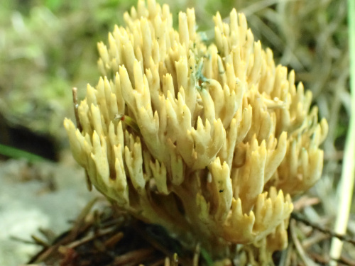
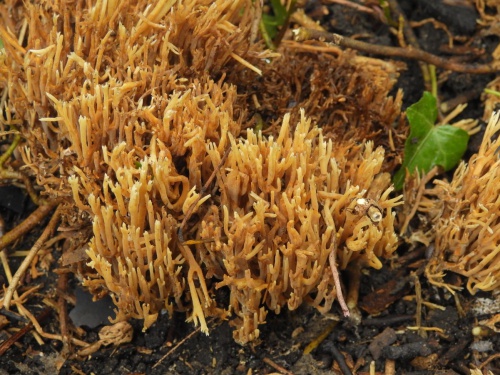
Gomphaceae - Corals
There are a large number of Ramaria species, and they are difficult to identify; microscopic examination and confirmation by an expert is usually needed. Growth form is usually densely and compactly branched, and they are either mycorrhizal or wood decomposers, associated with roots, woody debris or woodchip, under trees or in woodland or scrub. Most start out pale yellow but become darker wth age. Some stain reddish-brown when bruised. Spore colour is described as cinnamon-buff, and as spores tend to collect in the branch forks of mature specimens, the middle part becomes a darker brown than the tips, which may remain pale. The spores are elliptic and warty. All Ramaria stain green when Ferrous sulphate (FeSO4) is applied to the hymenium (the fertile surface). Some have a distinctive smell and taste, so this should always be noted.
Phaeoclavulina have now been separated from Ramaria, but may appear under this name in some sources. They usually have greenish, brownish or curry-yellow colours, and some stain green when bruised. Spores are teardrop-shaped with spiny warts. The fruitbody surface also stains green with FeSO4.