All images on this website have been taken in Leicestershire and Rutland by NatureSpot members. We welcome new contributions - just register and use the Submit Records form to post your photos. Click on any image below to visit the species page. The RED / AMBER / GREEN dots indicate how easy it is to identify the species - see our Identification Difficulty page for more information. A coloured rating followed by an exclamation mark denotes that different ID difficulties apply to either males and females or to the larvae - see the species page for more detail.
Fungi
Fungi are not plants, as was thought to be so in the past, but in a separate Kingdom of their own. In most cases, the main body of the fungus is hidden from view. It is a network of threads (hyphae) collectively called a 'mycelium', which permeate the substrate on which the fungus grows. The hyphae absorb nutrients and water from the substrate. The reproductive spore-producing structures, such as the familiar mushroom or bracket, are the visible parts of the fungus.
The Fungus Kingdom is extremely diverse, and includes many microfungi that are rarely recorded. Most of the larger species are in one of two major divisions or Phylum, based on the way the fungus reproduces. Basidiomycota (sometimes called 'spore-droppers') include mushrooms and toadstools, brackets, corals, puffballs, jellies, rusts and smuts. The Ascomycota or 'spore-shooters' includes cups, morels, yeasts and many other smaller fungi.
Taxonomy is very complicated and is liable to change as the true relationships between species are worked out. In the sections below, we have grouped fungi into categories based on their structure and appearance rather than their place in the scientific taxonomic hierarchy. There is more information under each section.
Recording fungi
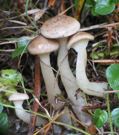
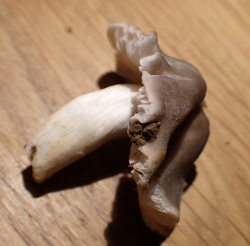
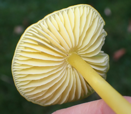
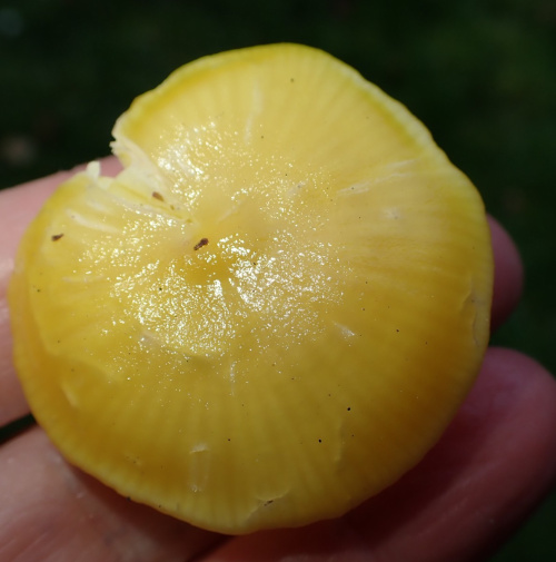
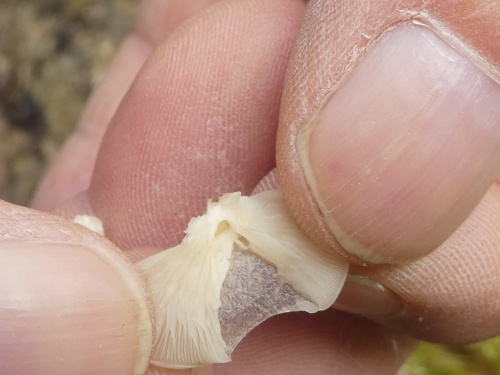
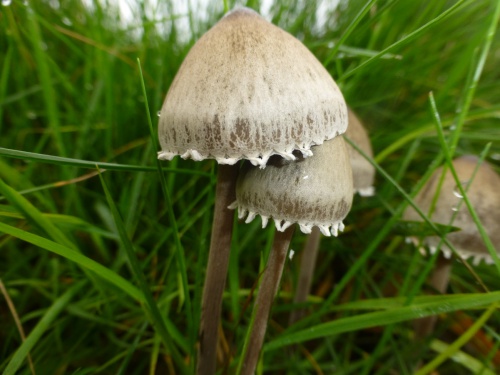
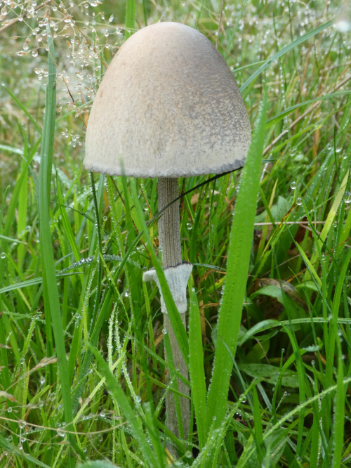
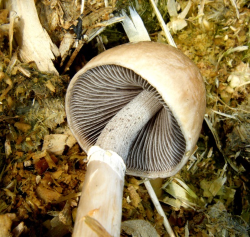
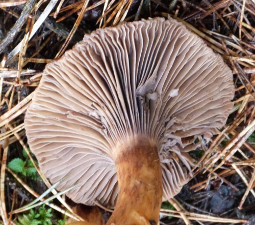
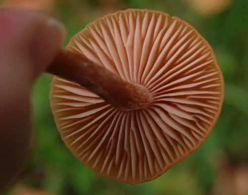
Always note the substrate or host-plant on which the fungus is growing, and the habitat. To identify many large species, it is helpful to get a good photograph of the cap (from the top), the gills or spore-producing structures (from underneath) and the stipe or stem (from the side) - three images from three angles, rather than images from much the same angle.
Always note the texture of cap and stipe; the smell; and the presence of any staining or 'milk' produced when the cap, gills, stipe or body of the fungus is bruised.
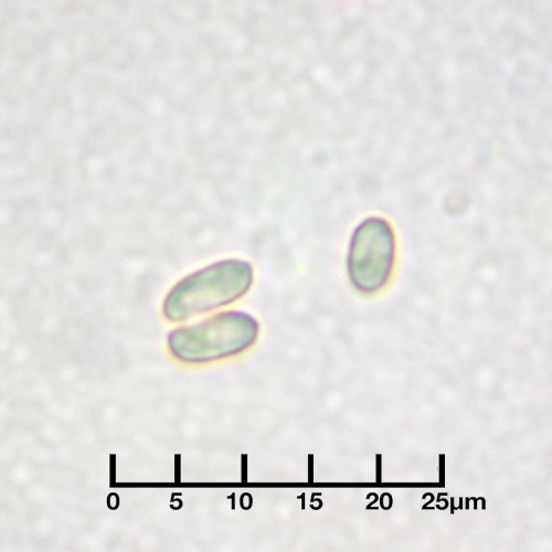
The colour of the spores can be very helpful. It is best seen by taking a spore-print from a collected specimen - advice on doing this is in many of the resources below. For some fungi, microscopic examination of the spores may be needed for identification.
Resources
The Leicestershire Fungi Study Group is a great place to start if you want to learn about fungi. They welcome new members. Their annual programme of study forays is led by enthusiastic and knowledgeable leaders. There are also indoor microscopy sessions held in Leicester. This is a really excellent way to broaden your knowledge, and make friends who share your interest in fungi.
There are many field guides, but none of them contain all the fungi you will find, or have all the photos or information you need, so it is a good idea to check several different books and websites.
- Kibby, G. 2020-2022. Mushrooms and Toadstools of Britain & Europe, Volumes 1 - 4. (privately published)
- Laessoe,T. & Petersen,J. (2019) Fungi of Temperate Europe. Princeton University Press, Volumes 1 and 2
- O'Reilly, P. (2016). Fascinated by Fungi. First Nature
- Sterry, P. & Hughes, B. (2009). Complete guide to British Mushrooms and Toadstools. Collins
- Wood, E & Dunkelman, J. (2017). Grassland Fungi: a Field Guide. Monmouthshire Meadows Group
Useful websites include Pat O'Reilly's First Nature website, the Buckinghamshire Fungus Group, the Hampshire Fungus Recording Group and Fungi Outer Hebrides.
The British Mycological Society website is a good source of information, mostly aimed at the expert.
If you know of other websites or books that you would recommend, do let us know: info@naturespot.org
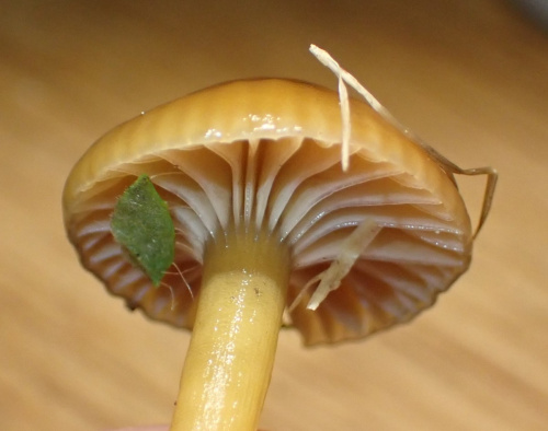
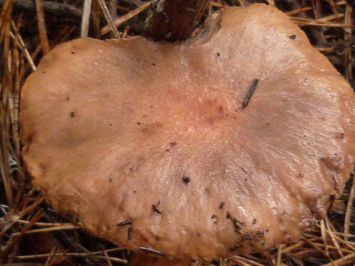
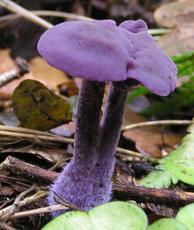
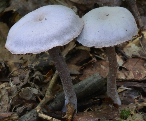
Agarics and other fungi with caps and gills
In this section are most of the commonly recorded 'mushrooms and toadstools' with spore bearing gills underneath the cap. Most of these are Agarics and related species, but we have included some families that have caps and gills but are unrelated - e.g. the Russulaceae.
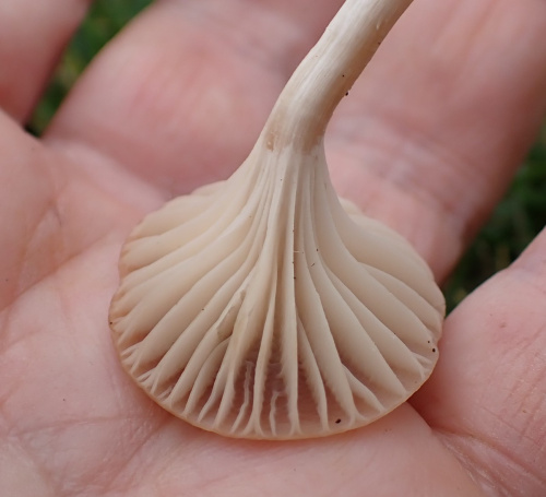
For most species, the spores fall from the gills and are dispersed by the wind. Spore colour is an important way of identifying species and it may be necessary to take a spore print from a collected specimen. As well as noting the colour, smell, texture, milk/staining, habitat and substrate, also note:
- the way the gills are attached to the stem or stipe; cutting vertically through the cap and stipe will help to see this (some examples are here: decurrent; adnate; adnexed/free)
- the colour of the gill edges (example: velvet shield)
- whether there is a ring around the stipe (some examples: wood mushroom; porcelain fungus)
- whether there is a volva or sac at the base of the stipe (example: false death-cap)
- whether there is a veil or the remains of one under the cap. (example: birch web-cap
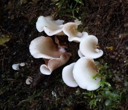
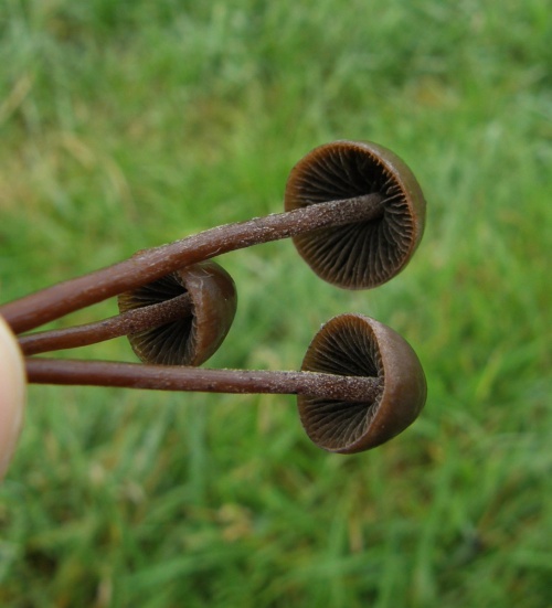
Crepidotaceae - Oysterlings
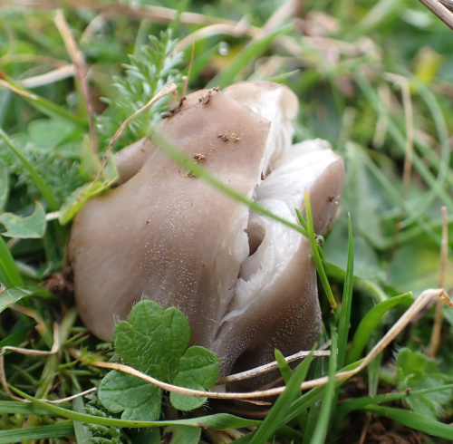
The Oysterlings (Crepidotus) are small fungi commonly found on twigs, branches and stumps of broadleaved deciduous trees. They have the unusual growth habitat of being attached to their substrate by the cap, often to one side, with the gills fanning out from the point of attachment. This growth habit is also seen in the Oyster mushrooms (Pleurotaceae), but these are larger and have pale spores, unlike the darker buff or brownish spores of Crepidotus. Identification of Crepidotus to species is difficult, usually requiring microscopic examination.
In the gallery below, an exclamation mark after the Red camera icon indicates that microscopic examination is needed.
The genus was formerly placed in the Inocybaceae.
 Crepidotus variabilis (Variable Oysterling) Leicester Road Quarry SP 4956 9341 (taken (12.11.2006)...JPG)
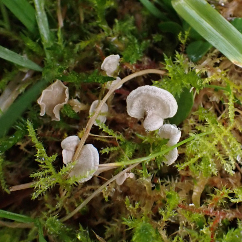
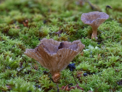
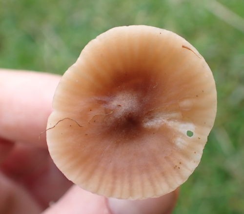
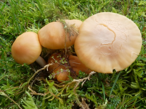
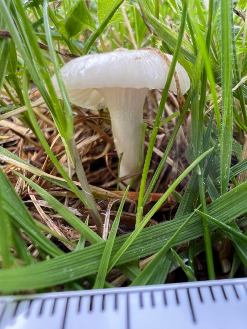
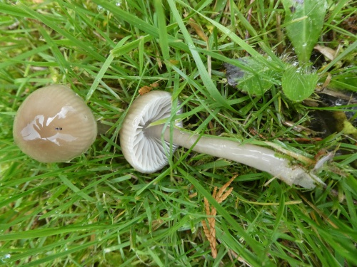
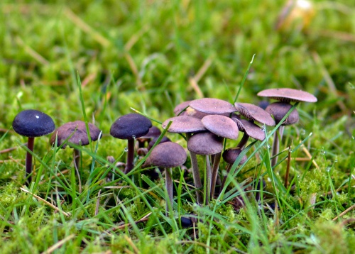
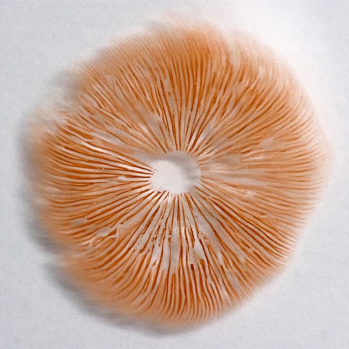
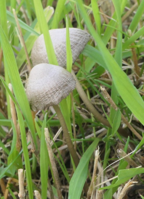
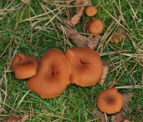
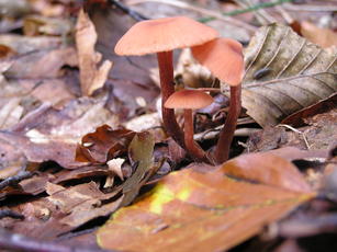
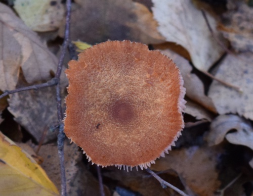
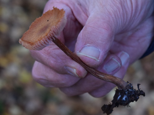
Entolomataceae - Pinkgills
The grassland Pinkgills are very difficult to identify, and microscopic examination of spores will almost always be needed. The genus can be recognised by the pinkish spores, see by taking a spore-print from a collected specimen. Most are grassland species, and along with waxcaps, clubs, coral and earth-tongues, they can indicate undisturbed grasslands of conservation value. Churchyards, cemeteries, old lawns and close-grazed nutrient-poor grasslands are good places to look.
In the gallery below, an exclamation mark after the Red or Amber camera icon indicates that microscopic examination or information on smell is needed
![]() The image is illustrative;
The image is illustrative;
identification not confirmed microscopically


![]() Pink spores. Illustrative image.
Pink spores. Illustrative image.
record not confirmed by microscopic
examination

 Entoloma conferendum (Star Pinkgill) Lawn of 39 Sharnford Road SP 4914 9316 (taken 14.11.2008),.JPG)
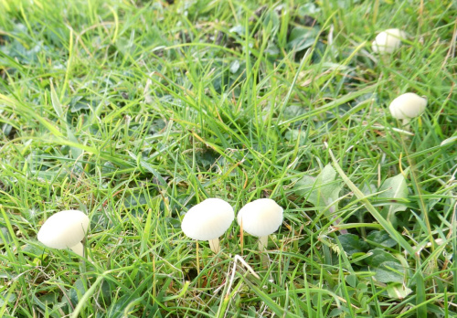
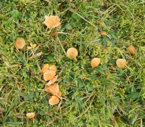
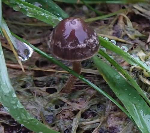
Galeropsidaceae - Mottlegills
The Mottlegills are associated with improved pastures with cattle, horse or sheep dung, or with lawns. As the name indicates, the gills are usually mottled blackish-brown when mature. They are hygrophanous - i.e. the cap changes colour depending on whether it is wet or dry; caps are darker in wet weather, becoming paler as they dry out. The Panaeolus spore print is black; Panaeolina is dark-brown.
The Dung Roundhead (Stropharia semiglobata) and some Deconica species are also associated with dung and improved grasslands and have dark spores; they can look very similar to Mottlegills; always check for the mottled gills. Conecaps (Conocybe) are also associated with dung, but have reddish brown spores.
In the gallery below, an exclamation mark after the Red camera icon indicates that microscopic examination is needed.

Gomphidiaceae - Spikes
Although we have included these species in this section because they have gills, not pores, they are actually more closely related to the Boletes and are in the order Boletales
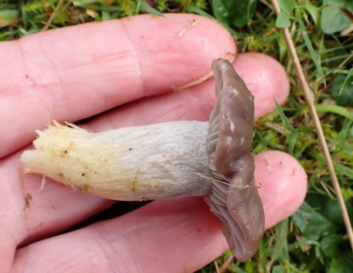
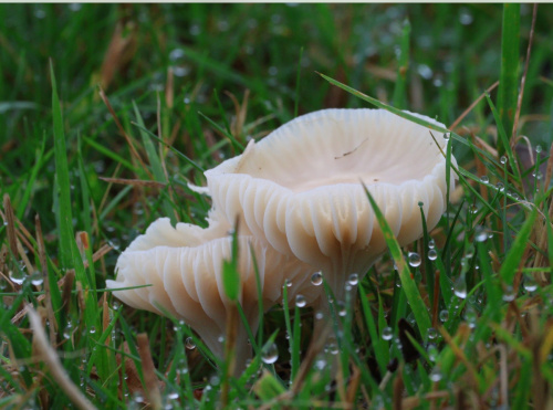
Hydnangiaceae - Deceivers
Deceivers have thick, waxy widely spaced gills, and white spiny spores.

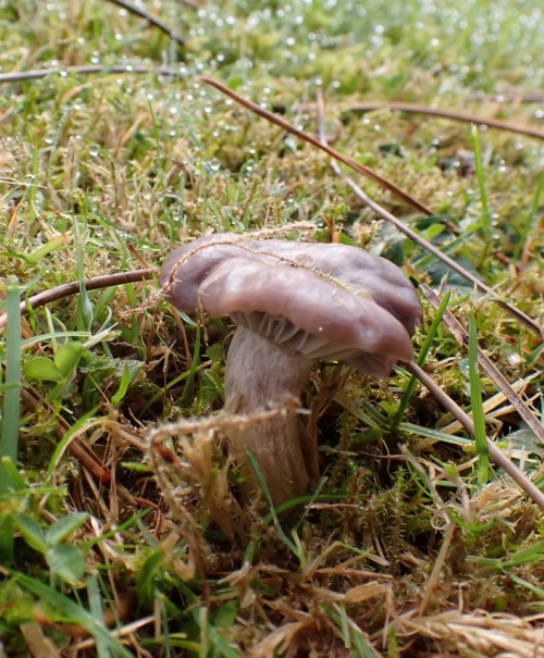
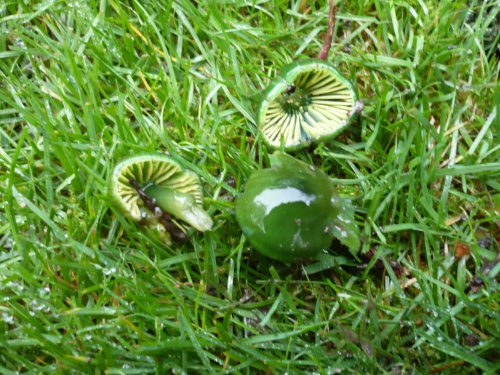
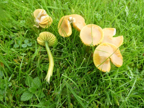
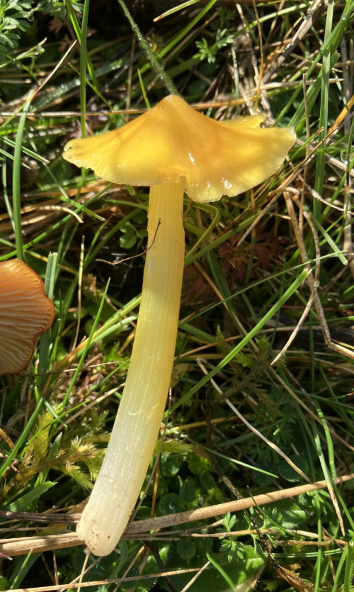
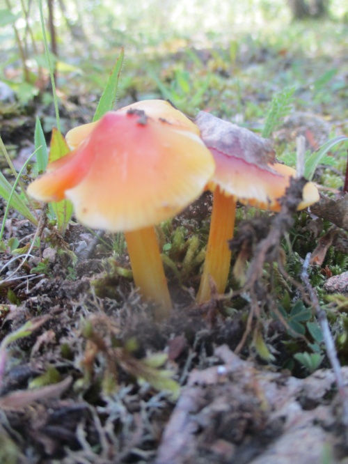
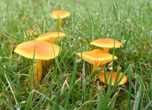
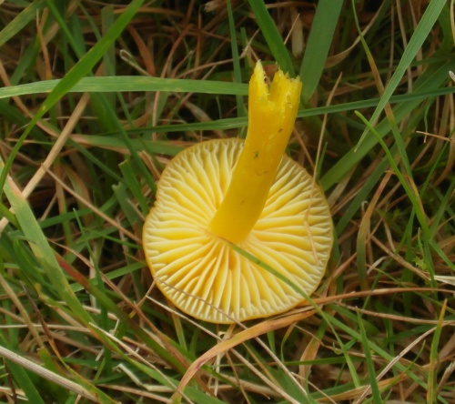
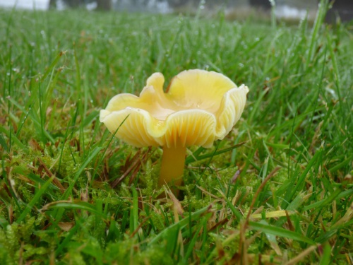
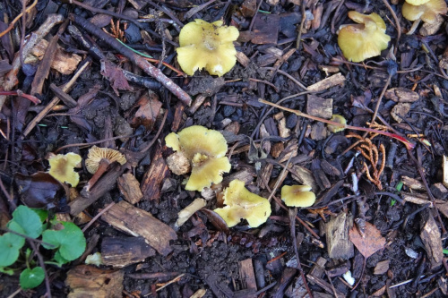
Hygrophoraceae - Waxcaps, Woodwaxes and Navels
Waxcaps are grassland fungi, favouring mown or closely grazed old unimproved grasslands, and have often been used as indicators of conservation-value grasslands. The Waxcap texture - slimy, dry, greasy or sticky (viscid) - of the cap and stipe are more important characters than colour, size or shape, which are very variable. The gills are waxy and widely spaced. Microscopic examination of the spores may help to identify some, but in general waxcap spores are all of similar size and shape, and are all white. Churchyards and cemeteries are good places to look. More information is here on the Leicestershire Fungi Study Group's website, and on these two helpful websites:
- The Grassland Waxcap Identification Tool by Claire Blencowe, for the Sussex BRC)
- The Waxcap Website (University of Wales - Aberystwyth)
In the gallery below, an exclamation mark after the Red or Amber camera icon for the Waxcap indicates that a note regarding smell, taste or texture is needed.