All images on this website have been taken in Leicestershire and Rutland by NatureSpot members. We welcome new contributions - just register and use the Submit Records form to post your photos. Click on any image below to visit the species page. The RED / AMBER / GREEN dots indicate how easy it is to identify the species - see our Identification Difficulty page for more information. A coloured rating followed by an exclamation mark denotes that different ID difficulties apply to either males and females or to the larvae - see the species page for more detail.
Fungi
Fungi are not plants, as was thought to be so in the past, but in a separate Kingdom of their own. In most cases, the main body of the fungus is hidden from view. It is a network of threads (hyphae) collectively called a 'mycelium', which permeate the substrate on which the fungus grows. The hyphae absorb nutrients and water from the substrate. The reproductive spore-producing structures, such as the familiar mushroom or bracket, are the visible parts of the fungus.
The Fungus Kingdom is extremely diverse, and includes many microfungi that are rarely recorded. Most of the larger species are in one of two major divisions or Phylum, based on the way the fungus reproduces. Basidiomycota (sometimes called 'spore-droppers') include mushrooms and toadstools, brackets, corals, puffballs, jellies, rusts and smuts. The Ascomycota or 'spore-shooters' includes cups, morels, yeasts and many other smaller fungi.
Taxonomy is very complicated and is liable to change as the true relationships between species are worked out. In the sections below, we have grouped fungi into categories based on their structure and appearance rather than their place in the scientific taxonomic hierarchy. There is more information under each section.
Recording fungi
Always note the substrate or host-plant on which the fungus is growing, and the habitat. To identify many large species, it is helpful to get a good photograph of the cap (from the top), the gills or spore-producing structures (from underneath) and the stipe or stem (from the side) - three images from three angles, rather than images from much the same angle.
Always note the texture of cap and stipe; the smell; and the presence of any staining or 'milk' produced when the cap, gills, stipe or body of the fungus is bruised.
The colour of the spores can be very helpful. It is best seen by taking a spore-print from a collected specimen - advice on doing this is in many of the resources below. For some fungi, microscopic examination of the spores may be needed for identification.
Resources
The Leicestershire Fungi Study Group is a great place to start if you want to learn about fungi. They welcome new members. Their annual programme of study forays is led by enthusiastic and knowledgeable leaders. There are also indoor microscopy sessions held in Leicester. This is a really excellent way to broaden your knowledge, and make friends who share your interest in fungi.
There are many field guides, but none of them contain all the fungi you will find, or have all the photos or information you need, so it is a good idea to check several different books and websites.
- Kibby, G. 2020-2022. Mushrooms and Toadstools of Britain & Europe, Volumes 1 - 4. (privately published)
- Laessoe,T. & Petersen,J. (2019) Fungi of Temperate Europe. Princeton University Press, Volumes 1 and 2
- O'Reilly, P. (2016). Fascinated by Fungi. First Nature
- Sterry, P. & Hughes, B. (2009). Complete guide to British Mushrooms and Toadstools. Collins
- Wood, E & Dunkelman, J. (2017). Grassland Fungi: a Field Guide. Monmouthshire Meadows Group
Useful websites include Pat O'Reilly's First Nature website, the Buckinghamshire Fungus Group, the Hampshire Fungus Recording Group and Fungi Outer Hebrides.
The British Mycological Society website is a good source of information, mostly aimed at the expert.
If you know of other websites or books that you would recommend, do let us know: info@naturespot.org
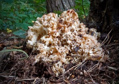
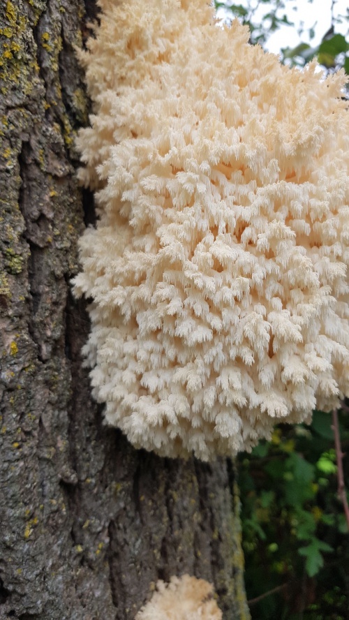
Polypores and Corticoids - brackets and crusts
This section includes many of the fungi that grow on the trunks, branches and stumps of trees. Often the species of tree on which they are growing - including dead wood - is an aid to identification. Almost all are decomposers of wood, causing white or brown rot.
Polypores release spores through downward pointing tubes on fertile surface. Pores can be labyrinthine (maze-like - see Oak Mazegill), circular, angular (e.g. Alder Bracket), elongated (e.g. Lumpy Bracket) or stretched and gill-like; always examine and photograph the pore layer. The number of pores per millimetre can be an important feature (use a transparent rule and hand lens). Some pore surfaces stain when bruised. Polypores can be perennial or annual; perennials e.g Artist's Bracket have several tube layers and often have zonate caps and hard or tough fruitbodies; annuals can be soft or tough and leathery. Polypores can have several different growth forms:
- Capped - a shelf/bracket or hoof-like form with a distinct cap on upper surface and a fertile surface with pores underneath e.g. Blushing Bracket or Hoof Fungus
- Resupinate – a flattened fruitbody without a cap, appressed to the substrate (e.g. Rusty Porecrust);
- Reflexed – partly resupinate, partly capped, with part of the fruit body bent back to form a bracket-like cap. e.g. Smoky Bracket;
- Clustered or fasciculate - numerous caps from a narrow common base or short thick stem e.g. Chicken-of-the-Woods, Dyer's Mazegill.
- Stemmed - tongue shaped with lateral stem attached to substrate (e.g. Birch Polypore or Dryad's Saddle), or with a central or acentric stem (e.g Winter Polypore and Bay Polpore). The latter may be superficially similar to Boletes, but polypore tubes cannot be separated from the cap flesh, unlike boletes
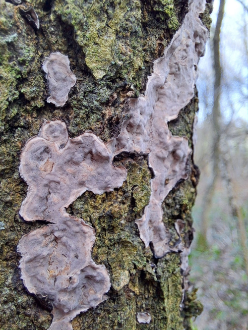
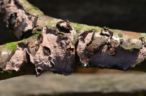
Corticoids do not have pores on the fertile surface and are usually resupinate or reflexed but may also be mould-like or fragile and loosely attached. Always photograph the fertile layer as well as the cap of reflexed forms. Some fertile surfaces stain when bruised e.g. Bleeding Oak Crust. The fertile surfaces can be wrinkled (merulioid) e.g. Wrinkled Crust or Netted Crust; smooth e.g. Hairy Curtain Crust, spiny or toothed e.g. Toothed Crust, gill-like e.g. Crimped Gill, or warty. Most are annual. Identification of many species is very difficult, and we have only illustrated a small number of UK species below. Microscopic examination by an expert will be needed to identify some.
Peniophoraceae - Crusts
Corticiaceae - crusts
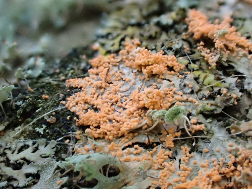
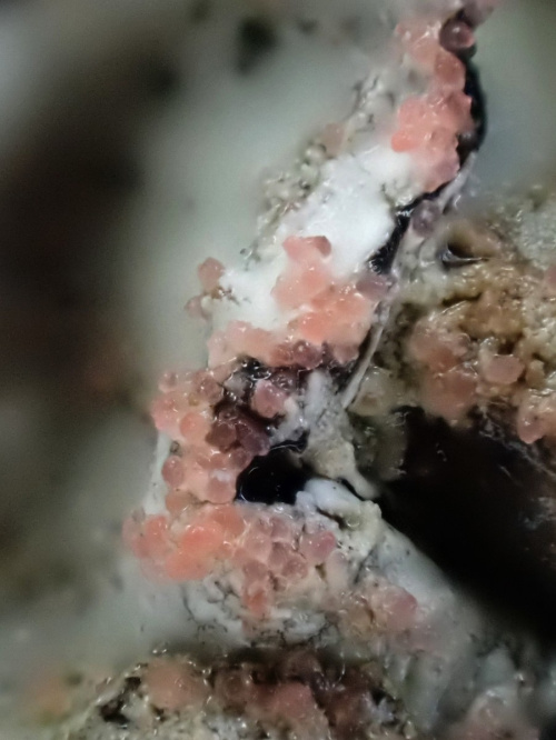
This family includes some lichenocolous fungi - i.e. those that live on and parasitise corticolous lichens such as Physcia and Xanthoria. You can find out more about this type of fungus on the British Lichen Society website. Also see Illosporiopsis christiansenii - this similar species is an ascomycete in a completely different part of the fungal kingdom.


Atheliaceae - Crusts
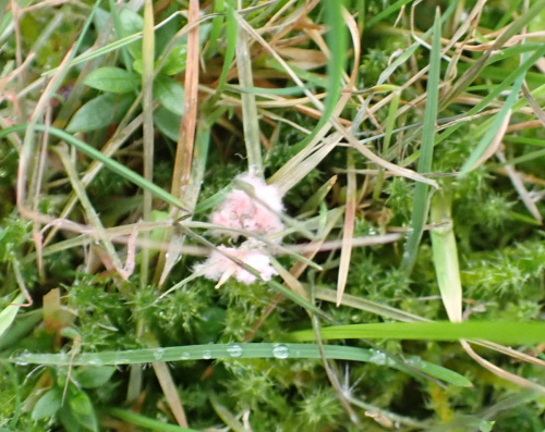
![]() cobwebby hyphae amongst
cobwebby hyphae amongst
the white thalli of the dead lichen
killed by the parasitic fungus

Botryobasidiaceae
Hydnodontaceae - crust
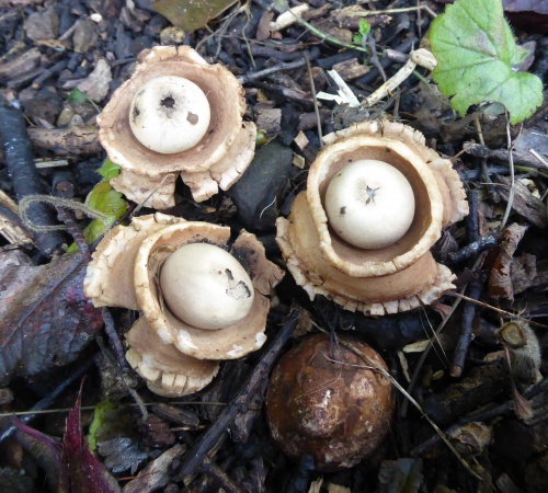
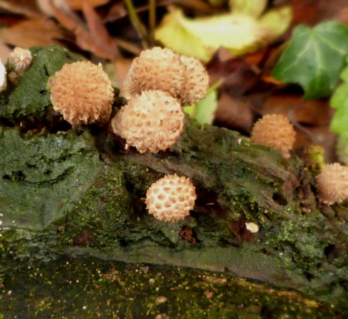
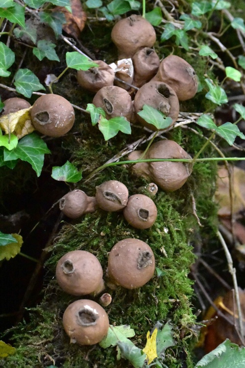
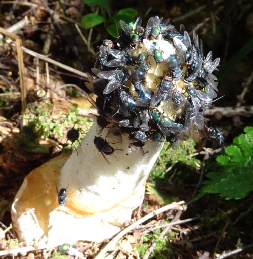
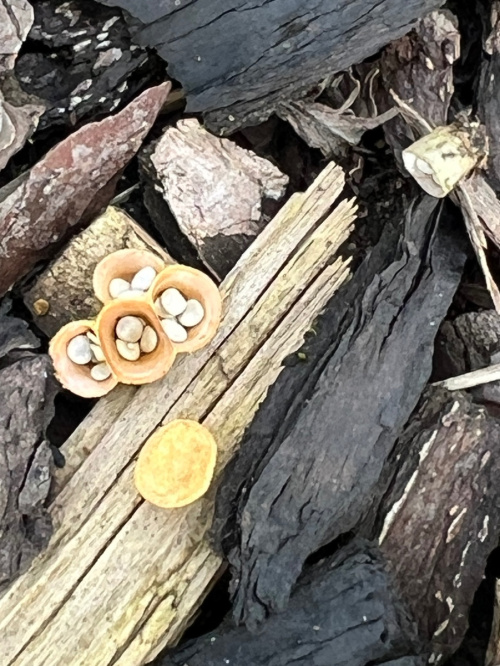
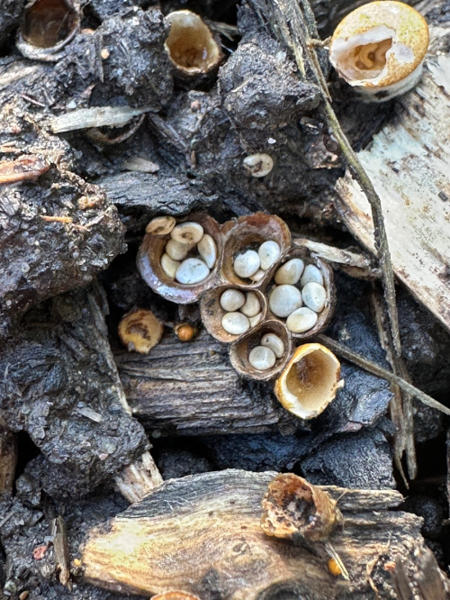
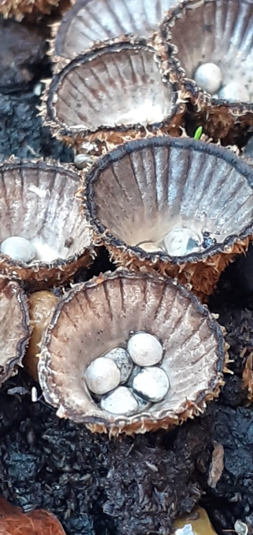
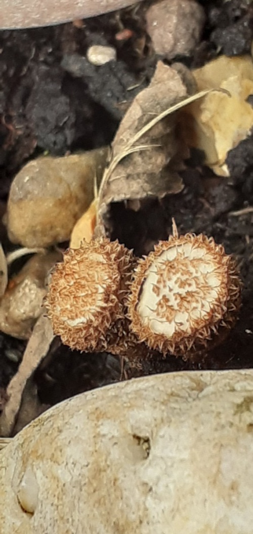
Bird's-nest Fungi
Despite their appearance, these tiny and often overlooked fungi are within the Agaricaceae, related to the Parasols and Field Mushrooms. They are found on twigs, rotten wood, decaying grass and vegetation, dung and woodchips, etc. Initially the cups are covered by a lid, or epiphragm, that falls away when the spores are mature. The spores are formed in egg-like structures called peridioles attached by a fine thread to the base of the tiny cup-shaped 'nest' or peridium. The peridioles are splashed out of the cup in heavy rain, snapping the thread and dispersing the 'eggs' several metres away.
Agaricacaeae - Bird's-nest fungi
Puffballs, Earthballs and Earth-stars
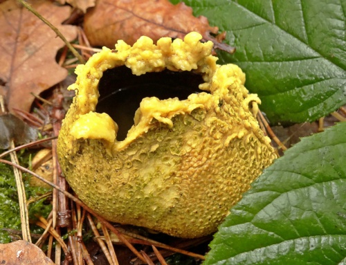
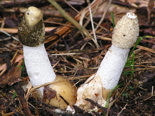
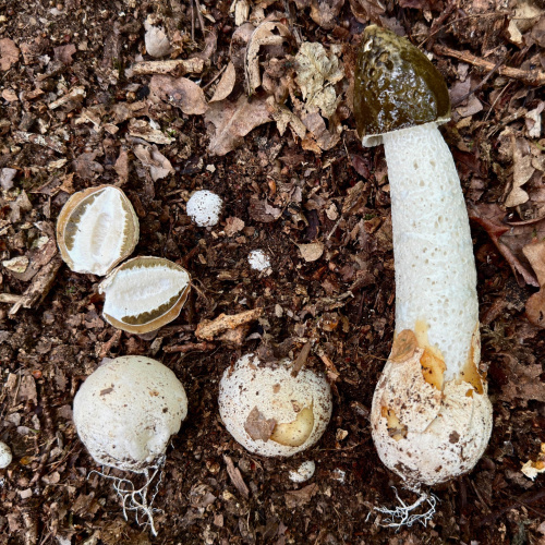
Sometimes called the gasteroid or stomach fungi, this group of species produces a powdery mass of spores inside the fruitbody, which opens via a central pore or ruptures when mature to release the spores which are then dispersed by rain or wind. Formerly they were grouped into an artifical family - Gasteromycetes - along with truffles, stinkhorns and bird's nest fungi, but it is now known that these species are not closely related.
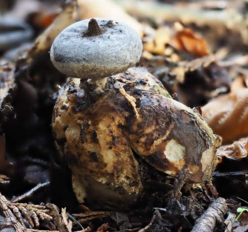
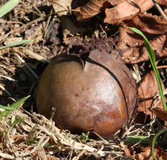
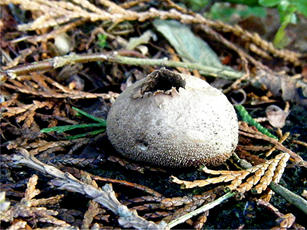
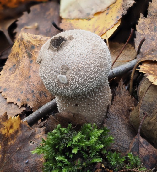
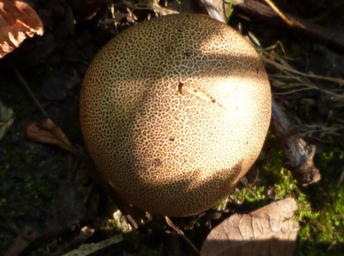
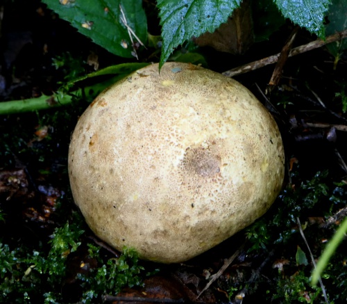
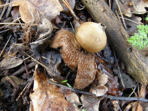
Earthballs (Scleroderma) have a thick, tough skin and a blackish marbled spore mass. They have a characteristic rubbery smell. The spores are dark, rounded and variously ornamented with a net-like reticulum, warts or short spines. When mature, the skin splits and the spores are released. Some have a root-like base and they tend to stay firmly attached to the substrate. Species are mycorrhizal and associated with a specific tree species or range of species, and they are found in woodland or under trees. They are more closely related to the Boletes than the Puffballs in Lycoperdaceae. See Malcolm Storey's article and key to British species in Field Mycology Vol 10 (4) 122-127 (2009), available on Science Direct here.
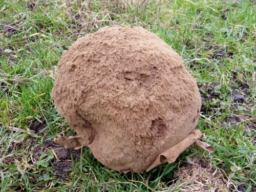
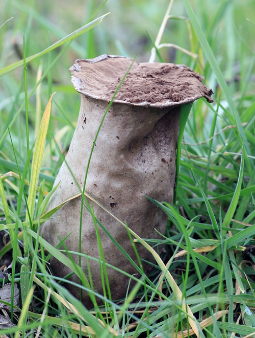
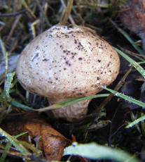
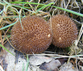
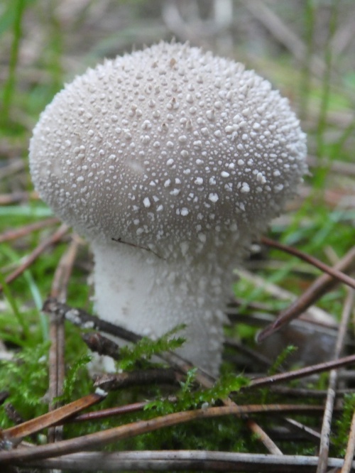
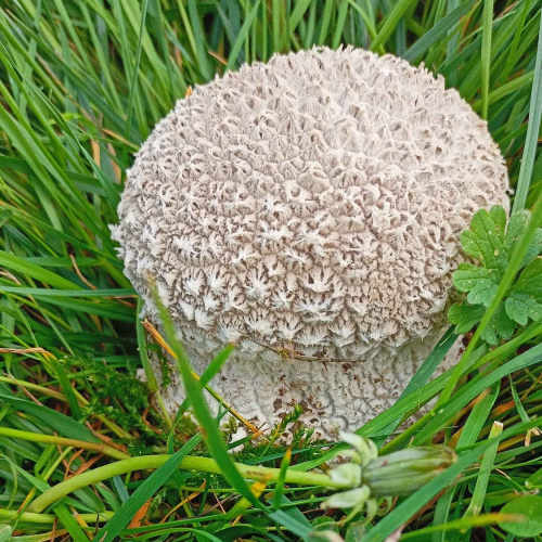
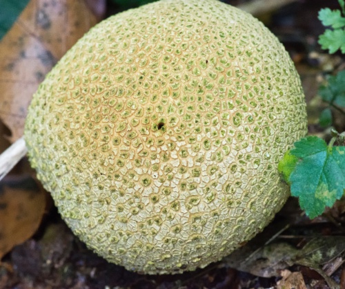
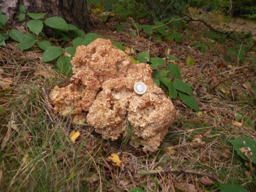
Puffballs (Lycoperdaceae) are a diverse family that include some large and impressive species. Some puffballs have spines or warts on the outer skin which are shed as it matures, leaving a mosaic-like pattern behind (e.g. Lycoperdon nigrescens, the Dusky Puffball). Some species release spores through a central pore (e.g. Lycoperdon and Bovista) and others disintegrate to leave a large spore-mass to disperse in wind and rain (e.g Calvatia, the Giant Puffball). Some species have a stem (e.g. the Pestle Puffball Lycoperdon excipuliforme). Bovista species have a thicker outer skin that cracks and falls away to reveal the thin inner skin around the spore-mass, which then becomes detached from the soil and rolls around to disperse the spores. Many are grassland species, but some may be found in woodland (e.g. the Common Puffball Lycoperdon perlatum) or associated with dead tree stumps or buried wood (e.g. the Stump Puffball Apioperdon pyriforme).
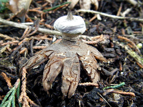
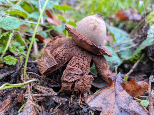
Earthstars (Geastrum) have an inner spore-sac surrounded by a thick outer skin. This cracks into segments or rays and peels away, raising the inner part up on the rays. Spores are eventually released from a central pore, which can be smooth, fringed or grooved and may resemble a beak, either within a clearly delimited zone or in a diffuse poorly defined area or halo. Some species have a stalk and a basal collar below the spore-sac (e.g. Geastrum striatum, the striated Earthstar). Some are hygroscopic - ie. the rays curl up when it is dry. The number of rays is variable. There is a key to European earthstars at https://www.mycokey.com/keys/FunDiveGeastrum.pdf
The Stalked Puffball (Tulostoma brumale) is very rare in Leicestershire and Rutland.
The Fenugreek Stalkball (Phleogena faginea) is a very small puffball, closely related to rusts, and is occasionally found on the bark of deciduous trees, especialy beech.
 Geastrum floriforme (Daisy Earthstar) Bassett Lane Sapcote SP 4872 9319 (taken 5.2.2010).JPG)