All images on this website have been taken in Leicestershire and Rutland by NatureSpot members. We welcome new contributions - just register and use the Submit Records form to post your photos. Click on any image below to visit the species page. The RED / AMBER / GREEN dots indicate how easy it is to identify the species - see our Identification Difficulty page for more information. A coloured rating followed by an exclamation mark denotes that different ID difficulties apply to either males and females or to the larvae - see the species page for more detail.
Fungi
Fungi are not plants, as was thought to be so in the past, but in a separate Kingdom of their own. In most cases, the main body of the fungus is hidden from view. It is a network of threads (hyphae) collectively called a 'mycelium', which permeate the substrate on which the fungus grows. The hyphae absorb nutrients and water from the substrate. The reproductive spore-producing structures, such as the familiar mushroom or bracket, are the visible parts of the fungus.
The Fungus Kingdom is extremely diverse, and includes many microfungi that are rarely recorded. Most of the larger species are in one of two major divisions or Phylum, based on the way the fungus reproduces. Basidiomycota (sometimes called 'spore-droppers') include mushrooms and toadstools, brackets, corals, puffballs, jellies, rusts and smuts. The Ascomycota or 'spore-shooters' includes cups, morels, yeasts and many other smaller fungi.
Taxonomy is very complicated and is liable to change as the true relationships between species are worked out. In the sections below, we have grouped fungi into categories based on their structure and appearance rather than their place in the scientific taxonomic hierarchy. There is more information under each section.
Recording fungi
Always note the substrate or host-plant on which the fungus is growing, and the habitat. To identify many large species, it is helpful to get a good photograph of the cap (from the top), the gills or spore-producing structures (from underneath) and the stipe or stem (from the side) - three images from three angles, rather than images from much the same angle.
Always note the texture of cap and stipe; the smell; and the presence of any staining or 'milk' produced when the cap, gills, stipe or body of the fungus is bruised.
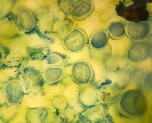
The colour of the spores can be very helpful. It is best seen by taking a spore-print from a collected specimen - advice on doing this is in many of the resources below. For some fungi, microscopic examination of the spores may be needed for identification.
Resources
The Leicestershire Fungi Study Group is a great place to start if you want to learn about fungi. They welcome new members. Their annual programme of study forays is led by enthusiastic and knowledgeable leaders. There are also indoor microscopy sessions held in Leicester. This is a really excellent way to broaden your knowledge, and make friends who share your interest in fungi.
There are many field guides, but none of them contain all the fungi you will find, or have all the photos or information you need, so it is a good idea to check several different books and websites.
- Kibby, G. 2020-2022. Mushrooms and Toadstools of Britain & Europe, Volumes 1 - 4. (privately published)
- Laessoe,T. & Petersen,J. (2019) Fungi of Temperate Europe. Princeton University Press, Volumes 1 and 2
- O'Reilly, P. (2016). Fascinated by Fungi. First Nature
- Sterry, P. & Hughes, B. (2009). Complete guide to British Mushrooms and Toadstools. Collins
- Wood, E & Dunkelman, J. (2017). Grassland Fungi: a Field Guide. Monmouthshire Meadows Group
Useful websites include Pat O'Reilly's First Nature website, the Buckinghamshire Fungus Group, the Hampshire Fungus Recording Group and Fungi Outer Hebrides.
The British Mycological Society website is a good source of information, mostly aimed at the expert.
If you know of other websites or books that you would recommend, do let us know: info@naturespot.org
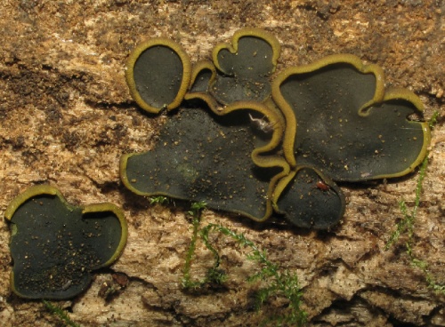
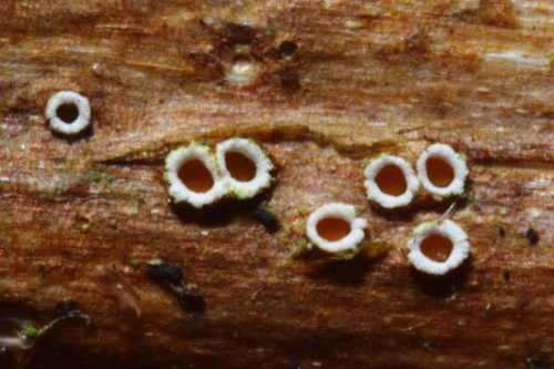
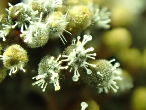
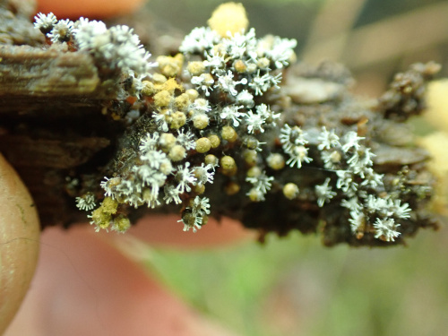
Jellies, Pins, Cups, Discos, etc.
This is an artifical grouping of species including those that form small disc or lens shaped fruit-bodies, jellies, pins or small cups. They are all ascomycetes, in various classes including Pezizomycetes, Leotiomycetes and Lecanoromycetes. Identification can be very difficult, and in the gallery below an exclamation mark after the Red camera icon idicates that microscopic examination of a specimen is needed.
Catinellaceae

Stictidaceae

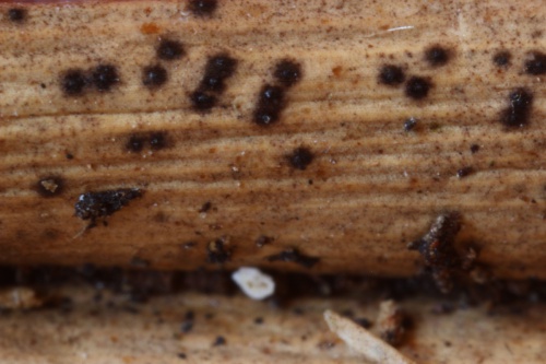
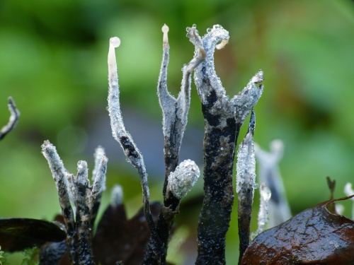
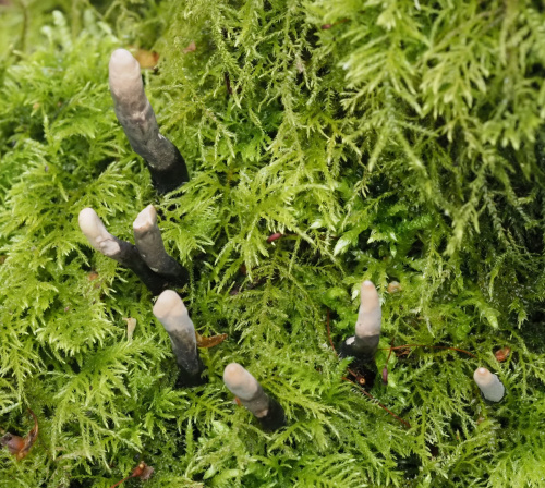
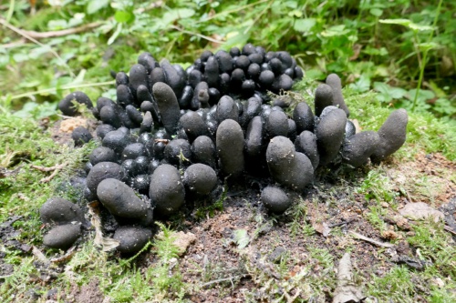
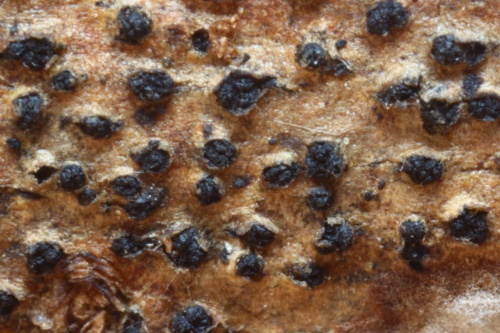
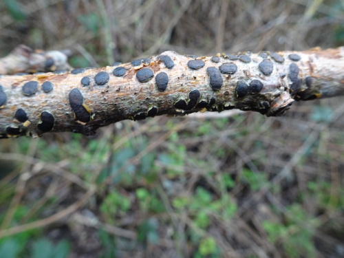
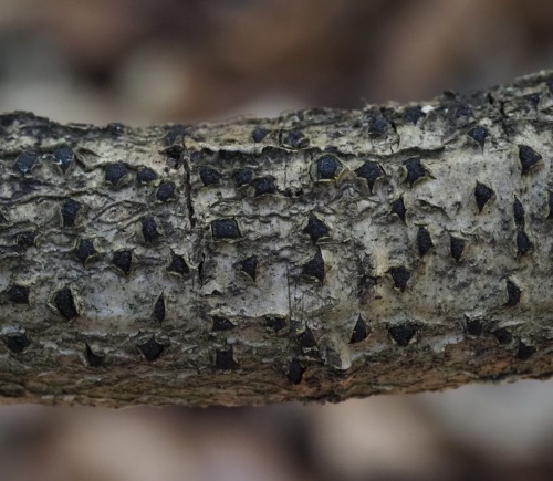
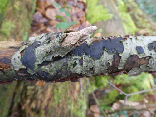
Snuffs, Fingers, Woodwarts & Barkspots
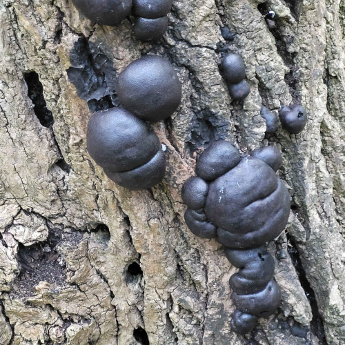
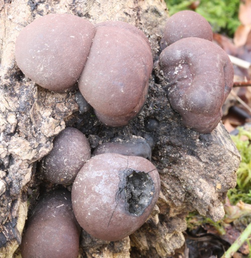
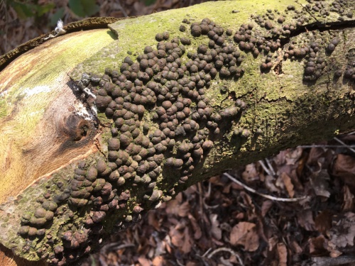
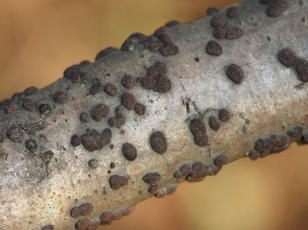
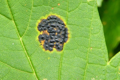
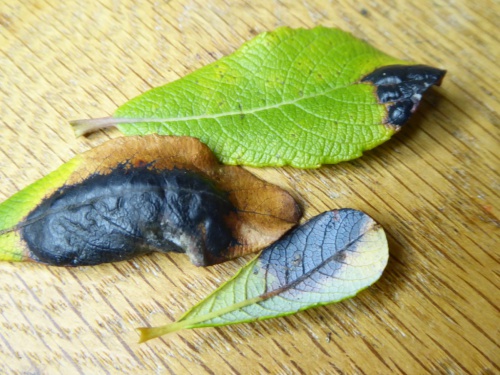
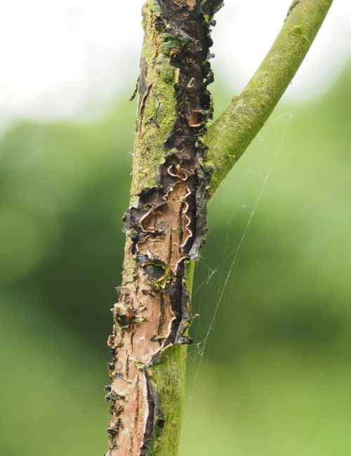
This group of species includes those that form hard black tar-like crusts or erumpent spots on bark; various other black or dark blisters, warts or pustules on bark or leaves and stems; and black fingers, balls and snuffs on rotting wood. They are all ascomycetes, most in the class Sordariomycetes.
Xylariaceae - Snuffs, Fingers


Diatrypaceae - Barkspots
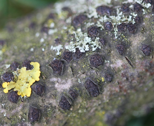
Hypoxylaceae - Woodwarts
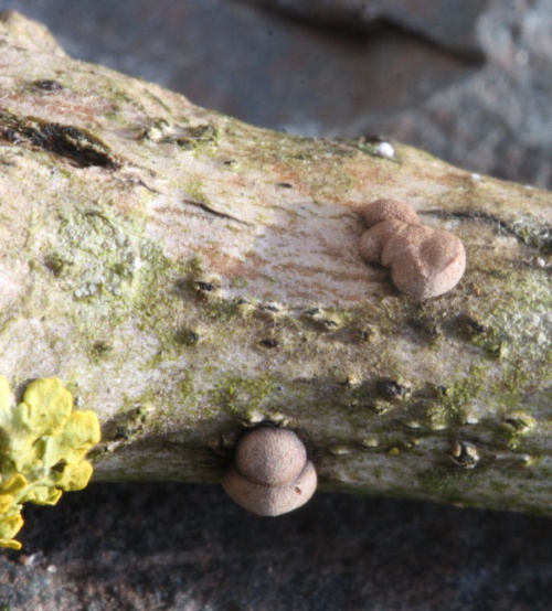
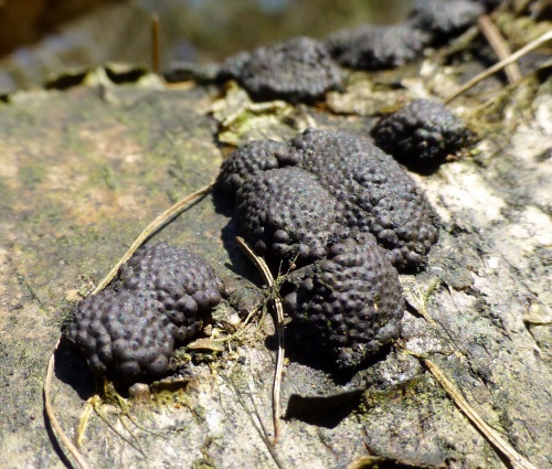
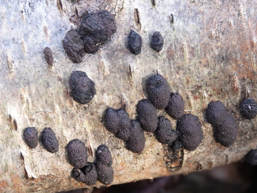
Graphostromataceae - Tarcrusts
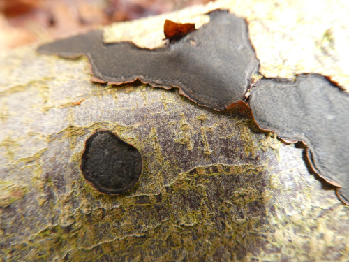
Rhytismataceae - Tarspots

Cryptomycetaceae - Blisters
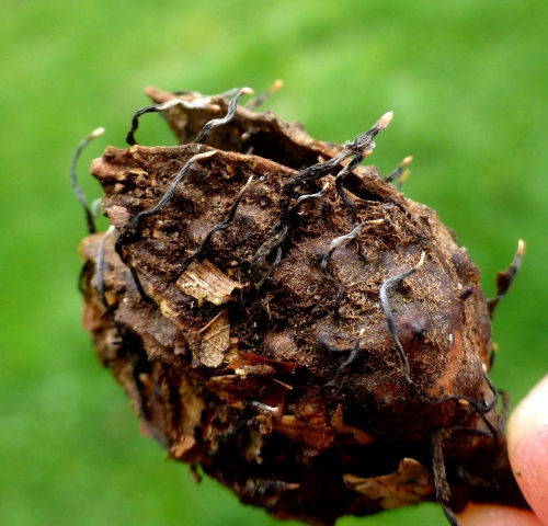
Caterpillarclubs; insect and slime mould pathogens
Cordycipitaceae
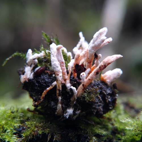
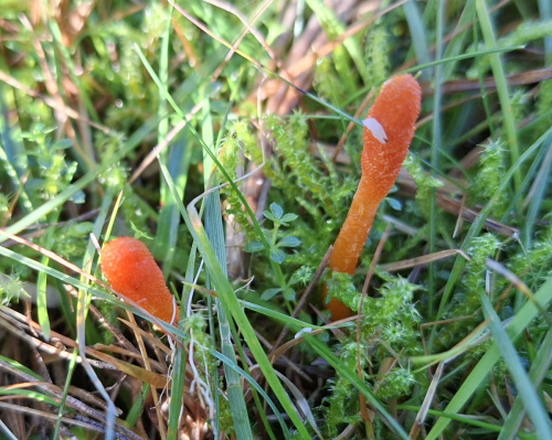
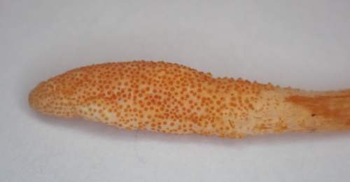
Ophiocordycipitaceae
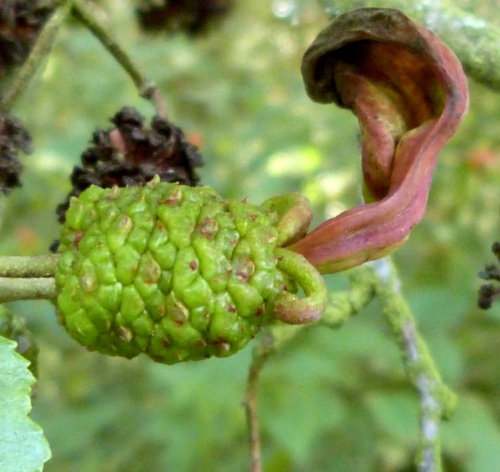
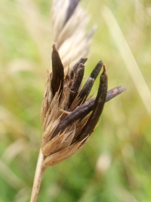
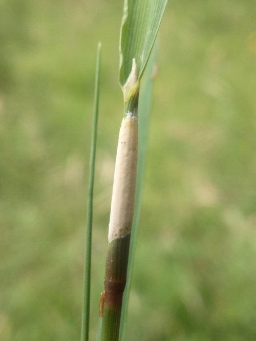
Choke and Ergot
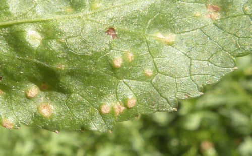
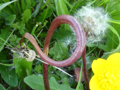
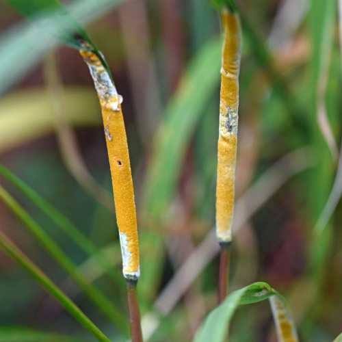
Ergots are elongate purplish-black galls that project from the individual florets of many species of grass and Spike-rushes.
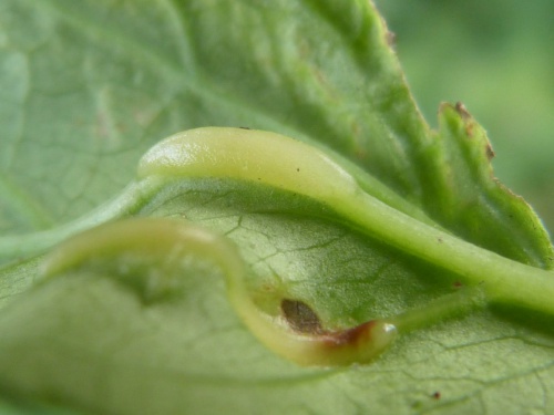
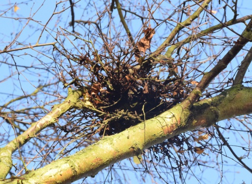
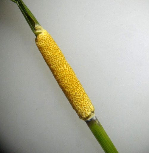
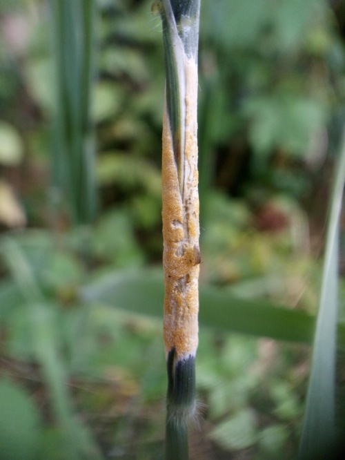
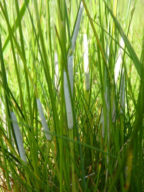
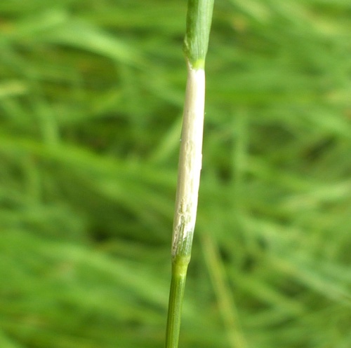
Chokes are galls formed by a conspicuous white or yellow stroma forming a tube encircling a grass-stem, sometimes including several stem-nodes. This prevents flowering. The stroma contains the asci and long narrow ascospores. It is now known that there are several species of Choke, depending on the species of grass affected: refer to British Plant Galls (Redfern and Shirley, 2023) for details.
- Agrostis (Bent) hosts = Epichloe baconii
- Holcus (Yorkshire Fog/Creeping Soft Grass) hosts = Epichloe clarkii
- Brachypodium sylvaticum (Wood False-brome) host = Epichloe sylvatica
- Bromus (Bromes) hosts = Epichloe bromicola
- Festuca (Fescues) hosts = Epichloe festucae
- Other host grasses including Poa (Meadow-grass), Dactylis glomerata (Cocksfoot), Anthoxanthum odoratum (Sweet Vernal Grass), Arrhenatherum elatius (False-Oat-grass), Lolium (Rye-grass), Phleum (Timothy) = Epichloe typhina
Clavicipitaceae
 ergots on Phleum pratense grass Field near Donkey Lane Sapcote SP 4860 9293 (taken 30.8.2009).JPG)
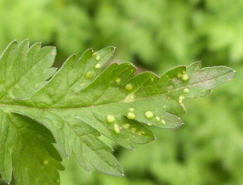
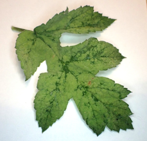
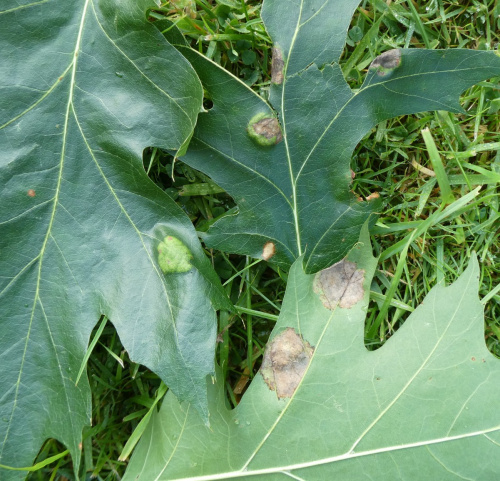
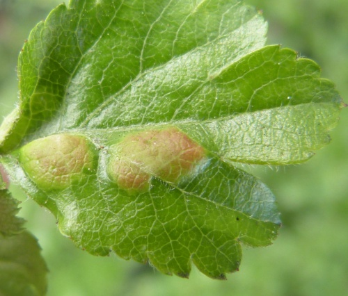
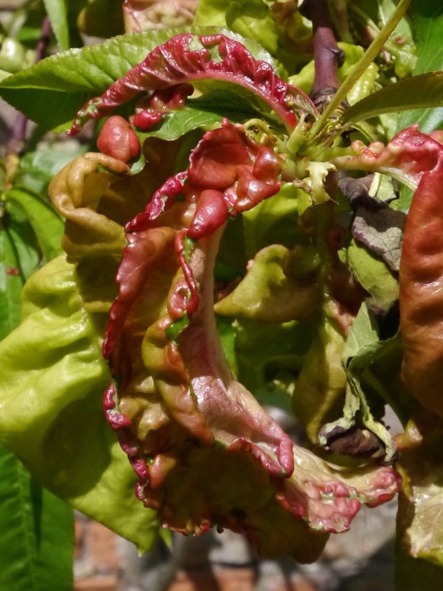
Gall fungi - Taphrinales
This order of fungi are Ascomycetes. Many species are gall-causers, and are fairly host-specific.
A variety of galls are formed by species in the genus Taphrina, such as deformed or 'pocket plum' fruit, witches' brooms, leaf curls or tongues. Typically, haploid Taphrina cells form yeast-like colonies early in their life-cycle. Cells germinate to produce invasive hyphae that infect new plant tissue. Eventually, a layer of asci containing the ascospores is produced on the leaves, fruits or other parts of the galled hosts, often in a visible bloom. Pressure builds up in the asci over summer, and they burst, shooting out the spores, which start the life cycle again if they land on a suitable host.